heart
[hahrt]Exercise stress testing is a valuable tool for detecting persons who have some degree of coronary heart disease. The test subject performs maximal exercise while being monitored by ECG. A positive stress test occurs when the subject cannot sustain the exercise for the duration of the test, cannot attain a normal maximal heart rate, or shows ECG changes indicative of ischemia. When stress testing is used for screening purposes, it is not diagnostic. However, persons with a positive stress test are 13 times more likely to develop significant coronary artery or heart disease and should work to reduce their risk factors. Stress testing is also used to evaluate the severity of known coronary disease and to guide the rehabilitation of a patient with coronary disease.
Phonocardiography is the recording of heart sounds and murmurs. It is more precise than auscultation with a stethoscope because it provides a permanent visual record that can be used to obtain precise timing information and can be used as baseline data for comparison with later findings.
Echocardiography is a type of diagnostic ultrasonography that provides information about the structure and function of the heart. It is a comfortable technique for the patient and is capable of establishing a diagnosis for several types of heart disease, especially those involving the valves. Types include M-mode, Doppler, and transesophageal echocardiography.
Several types of radioisotope examination are used to detect heart disease. A radioisotope imaging agent is injected into the patient, and a scintillation camera is then used to make an image of the distribution of radioactivity.
Thallium 201 has an affinity for heart tissue; when injected intravenously, it is carried to areas with adequate perfusion. Myocardial infarcts and areas of acute ischemia or scarring appear as “cold spots” (areas of no uptake of thallium) on the scintigram. When the isotope is injected during maximal exercise in an exercise stress test, the scan shows areas of inadequate perfusion and is a better indicator of coronary disease than a stress test alone.
Radiopharmaceuticals that label the blood pool can be used with a computerized scintillation camera to evaluate ventricular performance. Images of the first pass of the radioisotope through the heart can be used to determine the cardiac output and ejection fraction, the size of the ventricles, and regional wall motion.
The imaging agents used for bone scans, such as technetium 99m pyrophosphate or diphosphonate, also have an affinity for areas of acute ischemic tissue damage. “Hot spots” on the scintigram (areas of isotope uptake) show areas of acute infarction. The scan is usually negative by approximately 6 days after an infarction.
Cardiac catheterization is an invasive technique used when definitive data are required to decide whether heart disease should be treated medically, surgically, or through interventional cardiology techniques such as percutaneous transluminal angioplasty, stents, or valvuloplasty. A catheter is inserted into a vein or artery, usually the brachial artery or the femoral vein or artery, and guided into the heart. Tracings of the pressure pulses within the chambers during the heart cycle are obtained. Cardiac output, pulmonary artery pressures, the orifice area of valves, and the degree of left-to-right shunting can be determined.
Angiocardiography is the x-ray examination of the heart after injection of a radiopaque contrast medium through a catheter at various locations in the heart. The films show the size and motion of the heart chambers and can demonstrate aortic or mitral regurgitation. In coronary arteriography the contrast medium is injected through a catheter into the orifice of each coronary artery. The films show atherosclerotic obstructions of the arteries and are useful in planning coronary bypass surgery, percutaneous transluminal angioplasty, or stents.
All these contributions to the control and correction of cardiovascular diseases are important in the reduction of mortality rates and improvement in patients' quality of life. Nevertheless, it is also important for prevention that there be an improvement of the general public's awareness of the causes and risk factors of cardiac disorders. Major risk factors that can be avoided, modified, or corrected include cigarette smoking, elevated blood lipids, obesity, habitual dietary excesses, lack of exercise, hypertension, and excessive stress. Health professionals can promote reduction in the incidence of heart disease by educating the public about these risk factors and by encouraging active participation in preventive measures, particularly in those that involve changes in lifestyle.
heart
(hart), [TA]heart
(härt)heart
See Abiomed implantable heart, Athlete's heart, Baby Fae heart, Bleeding heart, Crisscross heart, Dextroposition heart, Depraved heart, Egg-shaped heart, Flabby heart, Flask-shaped heart, Holiday heart, Left heart, Mongolian heart, Old soldier's heart, Penn State heart, Right heart, Sabot heart, Second heart, Stone heart, Swinging heart, Water bottle heart.HEART
Cardiology A clinical trial–Healing & Early Afterload Reducing Therapyheart
(hahrt) [TA]Synonym(s): cor [TA] , coeur.
heart
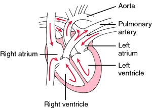
The twin-sided, four-chambered controlled muscular pump that, by means of regular rhythmical tightening (contractions) of the chambers and the action of valves, maintains the twin circulations of blood to the lungs and to the rest of the body. The right side of the heart pumps blood through the lungs and back to the left side. The left side pumps the blood returning from the lungs through all parts of the body and back to the right side.)
heart
the muscular pump of the BLOOD CIRCULATORY SYSTEM. In those invertebrates that possess a heart (e.g. ARTHROPODS, ANNELIDS, MOLLUSCS, ECHINODERMS) the heart is composed of several chambers and lies dorsal to the gut. In vertebrates the heart is made of special CARDIAC MUSCLE and lies in a ventral position surrounded by the PERICARDIUM. The five classes of vertebrates show an increasing complexity of structure, from the simple S-shaped heart with one ATRIUM and one VENTRICLE (2) found in fish, through the amphibians and most reptiles where the heart is divided into two atria but retains a single ventricle, and on to the birds and mammals where the heart shows complex separation into two sides with two atria and two ventricles. The main features of the human heart are:- the right side pumps blood around the pulmonary (lung) circulation for oxygenation, the left side pumping blood around the systemic (body) circulation where it becomes deoxygenated.
- blood from the body enters the right atrium via the superior vena cava (upper body) and inferior vena cava (lower body). A coronary sinus also drains into the right atrium bringing blood from the heart itself. The right atrium squeezes blood through the atrioventricular (AV) opening into the muscular right ventricle. finally, blood is ejected into the single opening of the pulmonary artery which splits to go to the two lungs.
- blood enters the left atrium from four pulmonary veins and passes through the left AV opening into the left ventricle. This has a much thicker wall than the right ventricle, reflecting its requirement for greater power. Blood leaves the left ventricle by one great vessel, the AORTA, which supplies all parts of the body, including the heart.
- flow of blood through the heart is in one direction only, due to the presence of various valves. Back-flow from ventricles to atria is prevented by AV valves, the tricuspid valve on the right side with three flaps, and the BICUSPID (3) valve on the left side with two flaps, both valves held in place by cords of connective tissue, the ‘chordae tendinae’. Back-flow from arteries to the ventricles is prevented by semilunar valves.
- various nerve areas connected with contraction are located in the heart (see HEART, CARDIAC CYCLE): (i) the sinoatrial node (SAN) or ‘pacemaker’ located in the wall of the right atrium near the entry of the venae cavae; (ii) the atrioventricular node (AVN) at the junction of all four heart chambers; (iii) the atrioventricular bundle, or bundle of His, running down the interventricular septum from the AVN; and (iv) a network of Purkinje tissue and other fibres spreading out from the bundle of His across the walls of both ventricles. See Fig. 189 .
Other nerve areas are situated in or near the heart: (i) baroreceptors in the walls of the heart, in the aortic arch, the carotid sinus, venae cavae and pulmonary veins where they enter the atria. Such sensory receptors are stimulated by stretching of the structure in which they are found, resulting in a decrease in blood pressure. (ii) chemoreceptors sensitive to blood CO2 levels are found in the AORTIC BODY and CAROTID BODY.
heart
(hahrt) [TA]Patient discussion about heart
Q. how does it feel to heart promblems answer to my question then talk to me
Other manifestations may include fainting (called syncope) either spontaneously or after exercise, edema (swelling) of the legs and various other non specific complaints.
The manifestations depend, of course, on the specific disease and the various characteristics of the patient (age, sex etc.)
You may read more here:
www.nlm.nih.gov/medlineplus/heartdiseases.html
Q. What happens to my heart when I exercise? My senior told me that exercise is good for health and especially for heart. What happens to my heart when I exercise?
Q. Is garlic helpful in heart ailments? I have heard that garlic is very good for cardiac health and using in curries or cooked with foods will be helpful. I have also heard that it has anti-inflammatory substances and also helps in weight loss. Is garlic helpful in heart ailments?
http://www.youtube.com/watch?v=_jOrw1eB-uc&eurl=http://www.imedix.com/health_community/vng-A24JmWJY_iceland_heart_protection_formula?q=heart&feature=player_embedded