Bundle Branch Block
Definition
Bundle branch block (BBB) is a disruption in the normal flow of electrical pulses that drive the heart beat.
Description
Bundle branch block belongs to a group of heart problems called intraventricular conduction defects (IVCD). There are two bundle branches, right and left. The right bundle carries nerve impulses that cause contraction of the right ventricle (the lower chamber of the heart) and the left bundle carries nerve impulses that cause contraction of the left ventricle. The two bundles initially are together at a junction called the bundle of His. Nerve impulses come through the sinus node of the heart to the bundle of His and then move into the right and left bundle branches. Bundle branch block is a slowing or interruption of nerve impulses. A problem may exist in any of the three bundles.
Patients with BBB are generally without symptoms unless the disease is severe enough to cause a complete infranodal A-V block and very slow heart rate. In patients with right bundle branch block (RBBB), the nerve impulse is conducted slowly or not at all. The right ventricle finally receives the impulse through muscle-to-muscle spread, outside the regular nerve pathway. This mechanism of impulse transmission is slow and results in a delayed contraction of the right ventricle. There are several types of left bundle branch block (LBBB), each producing its own characteristic mechanism of failure. In each case, the nerve impulse is blocked or delayed. Patients with LBBB may have left ventricular disease or cardiomyopathy.
Causes and symptoms
Left bundle branch block usually happens as a consequence of other diseases such as arteriosclerosis, rheumatic fever, congenital heart disease, myocarditis, myocardial infarction, metastatic heart tumors, or other invasions of the heart tissue. Right bundle branch block happens less often from underlying heart disease.
Diagnosis
Detection of BBB usually takes place during a normal physical examination. The block shows up as a widening of the second heart sound. Confirmation of BBB is obtained by electrocardiogram (ECG). The pattern seen in the electrocardiogram indicates pulses in a heart beat and their duration. A QRS duration of greater than 110 milliseconds is a diagnostic indication of BBB. There is a unique ECG pattern for blocks in each of the three bundles.
Treatment
There is no specific therapy for BBB. Patients are usually treated for associated heart diseases.
Prognosis
The prognosis of blockage in any of the three bundle branches depends on the prognosis of the associated heart disease. The associated diseases determine the outcome of the patient's health. Occasionally, disruptions in bundle branches lead to complete infranodal A-V block, a more serious blockage of nerve impulses. Approximately 2% of patients with BBB develop infranodal A-V blockage and these patients often require artificial pacemakers.
Resources
Books
Alexander, R. W., R. C. Schlant, and V. Fuster, editors. The Heart. 9th ed. New York: McGraw-Hill, 1998.
Key terms
Electrocardiogram — The pattern of the heart's electrical impulses that indicate the order and condition of the heart's components.
QRS — A pattern seen in an electrocardiogram that indicates the pulses in a heart beat and their duration. Variations from a normal QRS pattern indicate heart disease.
Gale Encyclopedia of Medicine. Copyright 2008 The Gale Group, Inc. All rights reserved.
block
[blok]1. to obstruct.
2. heart block.
atrioventricular block
1. any interruption of the conduction of electrical impulses from the atria to the ventricles; it can occur at the level of the atria, the atrioventricular node, the bundle of His, or the Purkinje system. See heart block.
2. a type of heart block in which the blocking is at the atrioventricular junction. It is first degree when atrioventricular conduction time is prolonged; it is called second degree or partial when some but not all atrial impulses reach the ventricle; and it is called third degree or complete when no atrial impulses at all reach the ventricle, so that the atria and ventricles act independently of each other.
atrioventricular block, complete see atrioventricular block.
atrioventricular block, first degree see atrioventricular block.
atrioventricular block, partial see atrioventricular block.
atrioventricular block, second degree see atrioventricular block.
atrioventricular block, third degree see atrioventricular block.
AV block atrioventricular block.
Bier block regional anesthesia by intravenous injection, used for surgical procedures on the forearm or the lower leg; performed in a bloodless field maintained by a pneumatic tourniquet that also prevents anesthetic from entering the systemic circulation. Called also intravenous block and IV block.
bifascicular block the combination of complete right bundle branch block with either left anterior fascicular block or left posterior fascicular block. This is an imprecise though commonly used term; specific terms defining the structures involved are preferred.
bundle branch block (BBB) a form of heart block involving delay or failure of conduction in one of the branches in the bundle of His, as determined by an electrocardiogram. It may be complete or incomplete, transient, permanent, or intermittent, and is also named according to involvement of the left or the right bundle branch. It is impossible to determine if bundle branch block is complete or not. When associated with acute anterior wall myocardial infarction, bundle branch block identifies a high-risk patient. See accompanying table.
bundle branch block, bilateral heart block characterized by conduction disturbance in the right and left bundle branches; it may be alternate, intermittent, or permanent. Complete bilateral bundle branch block results in complete (third degree) atrioventricular block.
bundle branch block, complete heart block characterized by absence of conduction in a bundle branch or conduction delay, causing ventricular activation to occur largely or exclusively through the contralateral bundle.
bundle branch block, incomplete heart block characterized by delayed conduction within a bundle branch, resulting in a delay in activation of the ipsilateral ventricle.
bundle branch block, right heart block characterized by a delay or failure of impulse propagation through the right bundle branch; it may be either complete or incomplete. See accompanying table.
caudal block caudal anesthesia.
cervical plexus block regional anesthesia of the neck by injection of a local anesthetic into the cervical plexus.
entrance block a zone of depressed conduction surrounding a pacemaker focus, protecting it from discharge by an extraneous impulse but not necessarily from discharges by electrotonic influences.
epidural block epidural anesthesia.
exit block heart block characterized by failure of an expected impulse to emerge from its focus of origin and propagate; this usually occurs with a parasystolic focus, but is also seen with sinus, junctional, and ventricular rhythms. In cardiac pacing it means that the pacemaker stimulus is not of sufficient amplitude to stimulate the heart, such as when there is a very high threshold.
fascicular block heart block characterized by certain abnormal QRS waveforms ascribed to conduction disturbance in the anterior and posterior divisions of the left bundle branch.
fascicular block, left anterior heart block characterized by delay or interruption of impulse conduction in the anterior superior division of the left bundle branch, resulting in asynchronous activation of the left ventricle.
fascicular block, left posterior heart block characterized by delay or interruption of impulse conduction in the posterior inferior division of the left bundle branch, resulting in asynchronous activation of the left ventricle.
femoral block regional anesthesia of the posterior thigh and the lower leg by injection of a local anesthetic around the femoral nerve just below the inguinal ligament at the lateral border of the fossa ovalis.
field block regional anesthesia by blocking conduction in nerves with chemical or physical agents.
heart block see heart block.
intravenous block Bier block.
intraventricular block impaired conduction within the ventricles due to absence of conduction within the bundle branches, their ramifications, or the ventricles.
intraventricular block, unspecified any heart block characterized by an electrocardiographic pattern of intraventricular conduction disturbance and not qualifying as a bundle branch block or a fascicular block.
interventricular block bundle branch block.
IV block Bier block.
lumbar plexus block regional anesthesia of the anterior and medial aspects of the lower limb by injection of a local anesthetic into the lumbar plexus.
mental block blocking (def. 3).
metabolic block the blocking of a biosynthetic pathway due to a genetic enzyme defect or to inhibition of an enzyme by a drug or other substance.
Mobitz type I block a second degree atrioventricular block in which the P-R interval increases progressively until an atrial impulse is blocked. Called also Wenckebach's phenomenon or block.
Mobitz type II block a second degree atrioventricular block in which the P-R interval is fixed, with periodic blocking of the atrial impulse to the ventricle.
nerve block regional anesthesia by injection of an anesthetic close to the appropriate nerve.
paracervical block regional anesthesia of the inferior hypogastric plexus and ganglia produced by injection of the local anesthetic into the lateral fornices of the vagina.
paraneural block perineural block.
parasacral block regional anesthesia by injection of a local anesthetic around the sacral nerves as they emerge from the sacral foramina.
paravertebral block paravertebral anesthesia.
perineural block regional anesthesia produced by injection of the anesthetic agent close to the nerve. Called also paraneural anesthesia or block and perineural anesthesia.
presacral block regional anesthesia produced by injection of the local anesthetic into the sacral nerves on the anterior aspect of the sacrum.
pudendal block regional anesthesia produced by blocking the pudendal nerves, accomplished by injection of the local anesthetic into the tuberosity of the ischium. 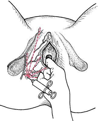

The pudendal block. The pudendal nerves can be effectively blocked by a local anesthetic, thereby anesthetizing the perineum. From Nichols and Zwelling, 1997.
retrobulbar block that performed by injection of a local anesthetic into the retrobulbar space to anesthetize and immobilize the eye.
sacral block regional anesthesia produced by injection of the local anesthetic into the extradural space of the spinal canal.
saddle block regional anesthesia in an area corresponding roughly with the areas of the buttocks, perineum, and inner aspects of the thighs, by introducing the anesthetic agent low in the dural sac. Called also saddle block anesthesia.
sinoatrial block a type of heart block characterized by partial or complete interference with the propagation of impulses from the sinoatrial node to the atria, resulting in delay or absence of the atrial response.
sinus block barosinusitis.
spinal block spinal anesthesia.
subarachnoid block spinal anesthesia (def. 2).
trifascicular block an imprecise term referring to heart block characterized by failure of conduction, partial or complete, in all three of the fascicles of the intraventricular conduction system; i.e., there is simultaneous right bundle branch block, left anterior hemiblock, and left posterior hemiblock. In the setting of acute anterior wall myocardial infarction, this is an ominous sign. More precise terms referring to the specifically involved structures are preferred.
vagal block (vagus nerve block) regional anesthesia produced by blocking of vagal impulses by injection of a solution of local anesthetic into the vagus nerve at its exit from the skull.
Wenckebach block Mobitz type I block.
wrist block regional anesthesia of the hand by injection of a local anesthetic around the median, radial, and ulnar nerves at the wrist.
Miller-Keane Encyclopedia and Dictionary of Medicine, Nursing, and Allied Health, Seventh Edition. © 2003 by Saunders, an imprint of Elsevier, Inc. All rights reserved.
bundle branch block
Cardiology Abnormal conduction through one of the conduction branches which normally supplies the right and left ventricles, often resulting in delayed conduction though either the right or left bundle branches. See Left bundle branch block, Right bundle branch block.McGraw-Hill Concise Dictionary of Modern Medicine. © 2002 by The McGraw-Hill Companies, Inc.
bundle branch block
Functional obstruction to one or other branch of the specialized muscle fibre conducting tissue of the heart which controls the timing of the contraction of the lower chambers (the ventricles). Bundle branch block usually implies that the heart muscle, generally, has been damaged, often by an inadequate blood supply from the coronary arteries. In extensive block, the outlook is poor.Collins Dictionary of Medicine © Robert M. Youngson 2004, 2005