glomerulonephritis
[glo-mer″u-lo-nĕ-fri´tis]a variety of nephritis characterized by inflammation of the capillary loops in the glomeruli of the kidney. It occurs in acute, subacute, and chronic forms and may be secondary to an infection, especially with the hemolytic streptococcus.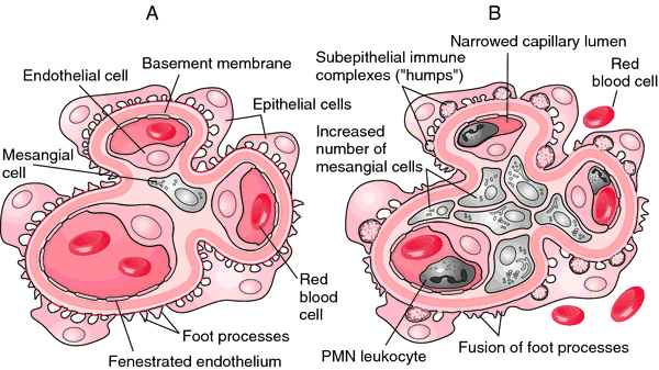

Histologic appearance of acute glomerulonephritis. A, Normal glomerulus. B, Glomerulonephritis. The glomerulus appears hypercellular and the capillaries are narrowed or occluded. From Damjanov, 2000.
diffuse glomerulonephritis a severe form of glomerulonephritis with proliferative changes in more than half the glomeruli, frequently with epithelial crescent formation and necrosis; it is often seen in cases of advanced systemic lupus erythematosus.
IgA glomerulonephritis IgA nephropathy.
lobular glomerulonephritis (membranoproliferative glomerulonephritis) a chronic glomerulonephritis characterized by mesangial cell proliferation and irregular thickening of the glomerular capillary wall. There are two subtypes: Type I is marked by subendothelial deposits and activation of the classic complement pathway. Type II is marked by heavy deposits in the glomerular basement membrane and activation of the alternative complement pathway. Both types occur in older children and young adults and follow a slowly progressing course with irregular remissions ultimately resulting in renal failure.
membranous glomerulonephritis a form characterized by proteinaceous deposits on the glomerular capillary basement membrane or by thickening of the membrane, with circulating antigen-antibody complexes indicating immune complex disease; it may be secondary to any of numerous other conditions. In some cases it may develop into the nephrotic syndrome. Called also membranous nephropathy.
mesangiocapillary glomerulonephritis membranoproliferative glomerulonephritis.
rapidly progressive glomerulonephritis acute glomerulonephritis marked by a rapid progression to end-stage renal disease and histologically by profuse epithelial proliferation, often with epithelial crescents; principal signs are anuria, proteinuria, hematuria, and anemia. Plasmapheresis or high doses of corticosteroids may lead to recovery of renal function.
Miller-Keane Encyclopedia and Dictionary of Medicine, Nursing, and Allied Health, Seventh Edition. © 2003 by Saunders, an imprint of Elsevier, Inc. All rights reserved.
mem·bra·no·pro·lif·er·a·tive glo·mer·u·lo·ne·phri·tis
chronic glomerulonephritis characterized by mesangial cell proliferation, increased lobular separation of glomeruli, thickening of glomerular capillary walls and increased mesangial matrix, and low serum levels of complement; occurs mainly in older children, with a variably slow progressive course, episodes of hematuria or edema, and hypertension. It is classified into three types: type 1, the most common, in which there are subendothelial electron-dense deposits; type 2, dense-deposit disease, in which the lamina densa is greatly thickened by extremely electron-dense material; type 3, in which there are both subendothelial and subepithelial deposits.
Synonym(s): hypocomplementemic glomerulonephritis, lobular glomerulonephritis, mesangiocapillary glomerulonephritis
Farlex Partner Medical Dictionary © Farlex 2012
membranoproliferative glomerulonephritis
A descriptive term for a pattern of diffuse glomerulonephritis (GN) characterised by mesangial cell proliferation and basement membrane reduplication. MPGN accounts for 5–10% of all GN; it is caused by immune deposits in glomeruli, and often progresses to chronic renal failure.Aetiology
Primary, typically linked to HCV-induced cryoglobulinaemia, or secondary to various infections including infectious endocarditis, malaria or in HCV infection due to intraglomerular deposition of immune complexes containing HCV, anti-HCV IgG, and IgM rheumatoid factors.
Clinical findings
Presentation ranges from nephrotic and nephritic syndromes to asymptomatic proteinuria and decreased serum C3.
Management
Symptomatic; also treat underlying disease.
Types
• Type I—60–70% of cases; subendothelial electron-dense material composed of C3, IgG, early complement components (C1q, C4, ±IgM).
• Type II—Ribbon type intramembranous dense deposits of C3 in lamina densa of glomerular basement membrane; also mesangial proliferation, crescentic GN.
• Type III—Extremely rare; subendothelial (type-I) and subepithelial immune deposits.
Segen's Medical Dictionary. © 2012 Farlex, Inc. All rights reserved.