effusion
[ĕ-fu´zhun]1. escape of a fluid into a part; exudation or transudation.
2. an exudate or transudate.
chyliform effusion see chylothorax.
chylous effusion see chylothorax.
pericardial effusion the accumulation of an abnormally large amount of pericardial fluid in the pericardium.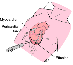

Accumulated fluid from a pericardial effusion evacuated by the subxiphoid approach to pericardiocentesis. From Polaski and Tatro, 1996.
pleural effusion see pleural effusion.
pseudochylous effusion see chylothorax.
Miller-Keane Encyclopedia and Dictionary of Medicine, Nursing, and Allied Health, Seventh Edition. © 2003 by Saunders, an imprint of Elsevier, Inc. All rights reserved.
per·i·car·di·al ef·fu·sion
increased fluid within the pericardial sac; can cause circulatory compromise by compression of the heart; most often caused by inflammation, infection, malignancy, and uremia.
Synonym(s): dropsy of pericardium
Farlex Partner Medical Dictionary © Farlex 2012
pericardial effusion
Medtalk An abnormal collection of fluid in the pericardiumMcGraw-Hill Concise Dictionary of Modern Medicine. © 2002 by The McGraw-Hill Companies, Inc.
per·i·car·di·al ef·fu·sion
(per'i-kahr'dē-ăl ĕ-fyū'zhŭn)Increased amounts of fluid within the pericardial sac, usually due to inflammation.
Medical Dictionary for the Health Professions and Nursing © Farlex 2012