macula
[mak´u-lah] (L.)1. a stain, spot, or thickening; in anatomy, an area distinguishable by color or otherwise from its surroundings. Often used alone to refer to the macula retinae.
2. a discolored spot on the skin that is not raised above the surface; called also macule.
3. a corneal scar that can be seen without special optical aids; it presents as a gray spot intermediate between a nebula and a leukoma.
4. macula lutea. adj., adj mac´ular, mac´ulate.
acoustic maculae (ma´culae acus´ticae) the macula sacculi and macula utriculi considered together.
macula atro´phica a white atrophic patch on the skin.
macula ceru´lea a blue patch on the skin seen in pediculosis.
macula cribro´sa a perforated spot or area; one of three perforated areas (inferior, medial, and superior) in the wall of the vestibule of the ear through which branches of the vestibulocochlear nerve pass to the saccule, utricle, and semicircular canals.
macula den´sa a zone of heavily nucleated cells in the distal renal tubule that feed information to the juxtaglomerular cells.
macula fla´va a yellow nodule at one end of a vocal cord.
macula folli´culi follicular stigma.
macula germinati´va germinal area; the part of the ovum where the embryo is formed.
macula lu´tea (macula lu´tea re´tinae) (macula re´tinae) an irregular yellowish depression on the retina, lateral to and slightly below the optic disk; receives and analyzes light only from the center of the visual field.
macula sac´culi a thickening on the wall of the saccule where the epithelium contains hair cells that receive and transmit vestibular impulses.
macula utri´culi a thickening in the wall of the utricle where the epithelium contains hair cells that are stimulated by linear acceleration and deceleration and by gravity.
Miller-Keane Encyclopedia and Dictionary of Medicine, Nursing, and Allied Health, Seventh Edition. © 2003 by Saunders, an imprint of Elsevier, Inc. All rights reserved.
mac·u·la
, pl.mac·u·lae
(mak'yū-lă, -ū-lē),1. A circumscribed flat area, up to 1 cm in diameter, differing perceptibly in color from the surrounding tissue.
See also: spot.
See also: neuroepithelium of macula.
See also: spot.
See also: neuroepithelium of macula.
2. A small discolored patch or spot on the skin, neither elevated above nor depressed below the skin's surface.
See also: spot.
See also: neuroepithelium of macula.
See also: spot.
See also: neuroepithelium of macula.
3. The neuroepithelial sensory receptors of the utricle and saccule of the vestibular labyrinth collectively.
See also: neuroepithelium of macula. Synonym(s): maculae utriculosaccularis [TA]
See also: neuroepithelium of macula. Synonym(s): maculae utriculosaccularis [TA]
[L. a spot]
Farlex Partner Medical Dictionary © Farlex 2012
macula
(măk′yə-lə)n. pl. macu·lae (-lē′) or macu·las
1.
a. An opaque spot on the cornea.
b. The macula lutea.
2. See macule2.
mac′u·lar adj.
The American Heritage® Medical Dictionary Copyright © 2007, 2004 by Houghton Mifflin Company. Published by Houghton Mifflin Company. All rights reserved.
macula
A circular area measuring 5 to 6 mm in diameter which is located on the posterior central retina. The macula has the fovea at its centre, which lies within the vacular arcades of the retina; it has a high concentration of photoreceptors, facilitates central vision and permits high-resolution visual acuity.Segen's Medical Dictionary. © 2012 Farlex, Inc. All rights reserved.
mac·u·la
, pl. maculae (mak'yū-lă, -lē)1. A small spot, perceptibly different in color from the surrounding tissue.
2. A small, discolored patch or spot on the skin, neither elevated above nor depressed below the skin's surface.
3. In ocular anatomy, indicates that portion of the retina located within the major vascular arcades, temporal to the optic nerve.
Synonym(s): macule, spot (1) .
Synonym(s): macule, spot (1) .
[L. a spot]
Medical Dictionary for the Health Professions and Nursing © Farlex 2012
macula
Any small flat spot.Collins Dictionary of Medicine © Robert M. Youngson 2004, 2005
macula
an area of acute vision on the retina of many vertebrates which lack a FOVEA.Collins Dictionary of Biology, 3rd ed. © W. G. Hale, V. A. Saunders, J. P. Margham 2005
Macula
The central part of the retina where the rods and cones are densest.
Mentioned in: Eye Examination
Gale Encyclopedia of Medicine. Copyright 2008 The Gale Group, Inc. All rights reserved.
fovea centralis
A small area of the retina of approximately 1.5 mm in diameter situated within the macula lutea. At the fovea centralis, the retina is the thinnest as there are no supporting fibres of Mueller, no ganglion cells and no bipolar cells. These cells are shifted to the edge of the depression. The fovea centralis contains mainly cone cells, each one being connected to only one ganglion cell and thus contributing to the highest visual acuity of the retina. The visual field represented by the fovea centralis is equal to about 5º (Fig. F9). Syn. foveal pit; macula (term often used by clinicians). See central visual acuity; retinal image; macula lutea.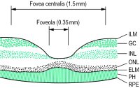

)
Fig. F9 Cross-section of the retina showing the fovea centralis and foveola (rod-free area)
macula lutea
An oval area of the retina 3-5 mm in diameter, with the foveal depression at its centre, slightly below the level of the optic disc and temporal to it (its centre lies 3.5 mm from the edge of the disc). The side wall of the depression slopes gradually towards the centre where the fovea centralis is located and where the best photopic visual acuity is obtained. Around the fovea, the ganglion cells are much more numerous than elsewhere, being arranged in five to seven layers. The outer molecular layer is also thicker than elsewhere and forms the outer fibre layer of Henle and there is a progressive disappearance of rods so that at the foveola only cones are found. The area of the macula lutea is impregnated by a yellow pigment (macular pigment) in the inner layers and for that reason is often called the yellow spot. Syn. area centralis (although that area is considered to be slightly larger, about 5.5 mm in diameter); punctum luteum. See blue field entoptoscope; fovea centralis; macular pigment.
Millodot: Dictionary of Optometry and Visual Science, 7th edition. © 2009 Butterworth-Heinemann
mac·u·la
, pl. maculae (mak'yū-lă, -lē)1. [TA] Circumscribed flat area, differing perceptibly in color from surrounding tissue.
2. Small discolored patch on skin, neither elevated above nor depressed below skin's surface.
3. Neuroepithelial sensory receptors of utricle and saccule of vestibular labyrinth collectively.
Synonym(s): spot (1) .
Synonym(s): spot (1) .
[L. a spot]
Medical Dictionary for the Dental Professions © Farlex 2012