Hydronephrosis
Definition
Hydronephrosis is the swelling of the kidneys when urine flow is obstructed in any of part of the urinary tract. Swelling of the ureter, which always accompanies hydronephrosis, is called hydroureter. Hydronephrosis implies that a ureter and the renal pelvis (the connection of the ureter to the kidney) are overfilled with urine.
Description
The kidneys filter urine out of the blood as a waste product. It collects in the renal pelvis and flows down the ureters into the bladder. The ureters are not simple tubes, but muscular passages that actively propel urine into the bladder. At their lower end is a valve (the ureterovesical junction) that prevents urine from flowing backward into the ureter. The bladder stores urine. The prostate gland surrounds the bladder outlet in males. Urine then flows through the urethra and out of the body as a waste product.
Because the urinary tract is closed save for the one opening at the bottom, urine cannot escape. Instead, the parts distend. Rupture is rare unless there is violent trauma like an automobile accident.
Obstructed flow anywhere along the drainage route can cause swelling of the upper urinary tract, but if the obstruction is below the bladder, the ureterovesical valve will protect the upper tract to a certain extent. Even then, with no place to go, the urine will back up all the way to its source. Eventually, the back pressure causes kidney function to deteriorate.
Obstruction need not be complete for problems to arise. Intermittent or partial obstruction is far more common than complete blockage, allowing time for the parts to enlarge gradually. Furthermore, if a ureterovesical valve is absent or incompetent, the pressure generated by bladder emptying will force urine backward into the ureter and kidney, causing dilation even without mechanical obstruction.
Causes and symptoms
Causes are numerous. Various congenital deformities of the ureter may sooner or later produce back pressure. Kidney stones are a common cause. They form in the renal pelvis and become lodged in the kidney, usually at the ureterovesical junction. In older men, the continued growth of the prostate gland leads commonly to restricted urine flow out of the bladder. Prostate cancer, and cancer anywhere else along the urine pathways, can obstruct flow. Pregnancy normally causes ureteral obstruction from the pressure of the enlarged uterus (womb) on the ureters.
Symptoms relate to the passage of urine. Sometimes, urine may be difficult to pass, irregular, or uncontrolled. Pain from distension of the structures is present. Blood in the urine may be visible, but it is usually microscopic.
In all cases where bodily fluids cannot flow freely, infection is inevitable. Symptoms of urinary infection may include:
- painful, burning urine
- cloudy urine
- pain in the back, flank, or groin
- fever, sweats, chills, and generalized discomfort
Patients often mistake a serious urinary infection for the flu.
Diagnosis
If the bladder is significantly distended, it can be felt through the abdomen. An analysis of the urine may reveal blood (if there is a stone), infection, or chemical changes suggesting kidney damage. Blood tests may also detect a decrease in kidney function.
All urinary obstructions will undergo imaging of some sort. Beginning with standard x rays to look for stones, radiologists, physicians specializing in the use of radiant energy for diagnostic purposes, will select from a wide array of tests. Ultrasound is simple, inexpensive, and very useful for these conditions. Standard x rays can be enhanced with contrast agents in several ways. If the kidneys are functioning, they will filter an x ray dye out of the blood and concentrate it in the urine, giving excellent pictures and also an assessment of kidney function. For better images of the lower urinary tract, contrast agents can be instilled from below. This is usually done with a cystoscope placed in the bladder. Through the cystoscope, a small tube can be threaded into the ureter through the ureterovesical valve, allowing dye to be injected all the way up to the kidney. CT and MRI scanning provide miraculous detail, more than is often needed for this condition.
Treatment
The obstruction must be relieved, even if it is partial or functional, as in the case of reflux from the bladder. If not, the kidney will ultimately be damaged, infection will appear, or both. The task may be as simple as placing a catheter through a restricting prostate or as complicated as removing a cancerous bladder and rebuilding a new one with a piece of bowel. In some cases, a badly damaged kidney may have to be removed.
Alternative treatment
Catheters or other urinary diversions may be better for weak or ill patients who cannot tolerate more extensive procedures. There is support using botanical medicine that can help the patient using a catheter avoid infections. Consultation with a trained health care practitioner is necessary.
Prognosis
After relief of the obstruction, a kidney may react with a brief flood of urine, but if the obstruction has been of short duration, normal kidney function will return. If one kidney is destroyed, the other will compensate for the lost organ.
Prevention
Kidney stones can be prevented by dietary changes and medication. Prompt evaluation of infections and urinary complaints will usually detect problems early enough to prevent long-term complications.
Resources
Organizations
American Association of Kidney Patients. 100 S. Ashley Dr., #280, Tampa, FL 33602. (800) 749-2257. http://www.aakp.org.
American Kidney Foundation. 6110 Executive Boulevard, #1010, Rockville, MD 20852. (800) 638-8299.
National Kidney Foundation. 30 East 33rd St., New York, NY 10016. (800) 622-9010. http://www.kidney.org.
Key terms
Catheter — A tube placed into the body that allows fluids to pass through it.
Contrast agent — Substances that cast shadows on x rays or other imaging methods.
CT and MRI — Two high technology methods of creating images of internal organs. Computerized axial tomography (CT or CAT) uses x rays, while magnetic resonance imaging (MRI) uses magnet fields and radio-frequency signals. Both construct images using a computer.
Cystoscope — A pencil-thin instrument that allows viewing and operating inside the urinary system.
Renal pelvis — The middle section of the kidney where urine first collects after filtration from the blood.
Gale Encyclopedia of Medicine. Copyright 2008 The Gale Group, Inc. All rights reserved.
hydronephrosis
[hi″dro-nĕ-fro´sis]distention of the renal pelvis and calices with urine; if it is allowed to progress, the functioning units of the kidney are destroyed. The collecting tubules dilate and the muscular walls of the pelvis and calices stretch, are replaced by fibrous tissue, and eventually form a large, fluid-filled, functionless sac. adj., adj hydronephrot´ic.
The cause of hydronephrosis is obstruction or atrophy of the urinary tract. Mechanical obstruction may result from ureteral tumors, calculi, benign or malignant hyperplasia of the prostate, or carcinoma of the bladder, urethra, or glans penis. Inflammatory obstruction is the outcome of a urinary tract infection that produces edema and narrowing of the ureters or urethra. Rarely there occurs during pregnancy a loss of muscle tone in the urinary tract. The atony is thought to be induced by placental hormones.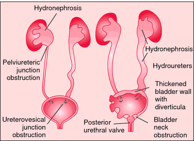
The cause of hydronephrosis is obstruction or atrophy of the urinary tract. Mechanical obstruction may result from ureteral tumors, calculi, benign or malignant hyperplasia of the prostate, or carcinoma of the bladder, urethra, or glans penis. Inflammatory obstruction is the outcome of a urinary tract infection that produces edema and narrowing of the ureters or urethra. Rarely there occurs during pregnancy a loss of muscle tone in the urinary tract. The atony is thought to be induced by placental hormones.
Symptoms. The patient usually complains of recurrent attacks of pain in the kidney region. The pain may be described as dull and nagging, or sharp. Examination of the urine often reveals the presence of pus and blood; there is fever if infection develops. If both kidneys are involved, uremia develops as the functional units of the kidneys are destroyed.
Diagnosis is established by extensive urologic examination with detailed pyelography, which usually reveals the cause of the obstruction and accumulation of fluid in the pelvis.
Diagnosis is established by extensive urologic examination with detailed pyelography, which usually reveals the cause of the obstruction and accumulation of fluid in the pelvis.
Treatment. The urinary tract must be drained by whatever means necessary; this may involve a simple dilatation of the ureter or urethra or it may require surgery of the affected kidney. When urinary tract infection is present as a cause or result of the hydronephrosis, urinary antiseptics and antibiotics are administered until the urine becomes sterile.

Obstruction to urine flow results in dilation of the urinary tract proximal to the site of obstruction. Obstruction may be at the pelviureteric or vesicoureteric junction (left), the bladder neck or urethra (right). From Lissauer and Young, 2002.
Miller-Keane Encyclopedia and Dictionary of Medicine, Nursing, and Allied Health, Seventh Edition. © 2003 by Saunders, an imprint of Elsevier, Inc. All rights reserved.
hy·dro·ne·phro·sis
(hī'drō-ne-frō'sis),Dilation of the pelvis and calyces of one or both kidneys. This may result from obstruction to the flow of urine, vesicoureteral reflux, or it may be a primary congenital deformity without an apparent cause.
Synonym(s): pelvocaliectasis, pyeloureterectasis
[hydro- + G. nephros, kidney, + -osis, condition]
Farlex Partner Medical Dictionary © Farlex 2012
hydronephrosis
(hī′drō-nə-frō′sĭs)n. pl. hydronephro·ses (-sēz)
The dilation of the pelvis and calyces of one or both kidneys because of the accumulation of urine resulting from obstruction of urine outflow.
The American Heritage® Medical Dictionary Copyright © 2007, 2004 by Houghton Mifflin Company. Published by Houghton Mifflin Company. All rights reserved.
hydronephrosis
Nephrology Uni- or bilateral expansion of a renal pelvis and calyces, often due to obstructive uropathy which may be linked to HTN and result in permanent renal damage. See Bilateral hydronephrosis, Reflux nephropathy, Unilateral hydrosis.McGraw-Hill Concise Dictionary of Modern Medicine. © 2002 by The McGraw-Hill Companies, Inc.
hy·dro·ne·phro·sis
(hī'drō-nĕ-frō'sis)Dilation of the pelvis and calyces of one or both kidneys resulting from obstruction to the flow of urine.
Synonym(s): pelvocaliectasis, uronephrosis.
Synonym(s): pelvocaliectasis, uronephrosis.
[hydro- + G. nephros, kidney, + -osis, condition]
Medical Dictionary for the Health Professions and Nursing © Farlex 2012
hydronephrosis
Ballooning out of the urine collecting system of the kidney, as a result of obstruction to the free outflow of urine at any point below the kidney. This may be due to simple or malignant prostate enlargement, external pressure on, or a stone or blood clot in, a URETER, tumour of the bladder, inflammatory narrowing of the URETHRA or even a very tight foreskin (phimosis). There is pain in the loin, and infection leads to fever and sometimes blood in the urine. Unrelieved hydronephrosis often proceeds to kidney failure.Collins Dictionary of Medicine © Robert M. Youngson 2004, 2005