nerve
[nerv]a macroscopic cordlike structure of the body, comprising a collection of nerve fibers that convey impulses between a part of the central nervous system and some other body region. See Appendix 2-6 and see color plates.
Depending on their function, nerves are known as sensory, motor, or mixed. Sensory nerves, sometimes called afferent nerves, carry information from the outside world, such as sensations of heat, cold, and pain, to the brain and spinal cord. Motor nerves, or efferent nerves, transmit impulses from the brain and spinal cord to the muscles. Mixed nerves are composed of both motor and sensory fibers, and transmit messages in both directions at once.
Together, the nerves make up the peripheral nervous system, as distinguished from the central nervous system (brain and spinal cord). There are 12 pairs of cranial nerves, which carry messages to and from the brain. Spinal nerves arise from the spinal cord and pass out between the vertebrae; there are 31 pairs, 8 cervical, 12 thoracic, 5 lumbar, 5 sacral, and 1 coccygeal. The various nerve fibers and cells that make up the autonomic nervous system innervate the glands, heart, blood vessels, and involuntary muscles of the internal organs.

Details of structure of components of nerve tissue.
accelerator n's the cardiac sympathetic nerves, which, when stimulated, accelerate the action of the heart.
acoustic nerve vestibulocochlear nerve; see anatomic Table of Nerves in the Appendices.
afferent nerve any nerve that transmits impulses from the periphery toward the central nervous system, such as a sensory nerve. See also neuron.
articular nerve any mixed peripheral nerve that supplies a joint and its associated structures.
auditory nerve vestibulocochlear nerve; see anatomic Table of Nerves in the Appendices.
autonomic nerve any nerve of the autonomic nervous system; called also visceral nerve.
cranial n's see cranial nerves.
cutaneous nerve any mixed peripheral nerve that supplies a region of the skin. See anatomic Table of Nerves in the Appendices.
depressor nerve
1. a nerve that lessens the activity of an organ.
2. an afferent nerve whose stimulation causes a fall in blood pressure.
efferent nerve any nerve that carries impulses from the central nervous system toward the periphery, such as a motor nerve. See also neuron.
excitor nerve one that transmits impulses resulting in an increase in functional activity.
excitoreflex nerve a visceral nerve that produces reflex action.
fusimotor n's those that innervate the intrafusal fibers of the muscle spindle.
gangliated nerve any nerve of the sympathetic nervous system.
inhibitory nerve one that transmits impulses resulting in a decrease in functional activity.
medullated nerve myelinated nerve.
mixed nerve (nerve of mixed fibers) a nerve composed of both sensory (afferent) and motor (efferent) fibers.
motor nerve a peripheral efferent nerve that stimulates muscle contraction.
myelinated nerve one whose axons are encased in a myelin sheath; called also medullated nerve.
peripheral nerve any nerve outside the central nervous system.
pilomotor n's those that supply the arrector muscles of hair.
pressor nerve an afferent nerve whose irritation stimulates a vasomotor center and increases intravascular tension.
sciatic nerve see sciatic nerve.
secretory nerve an efferent nerve whose stimulation increases vascular activity.
sensory nerve a peripheral nerve that conducts impulses from a sense organ to the spinal cord or brain. See also neuron.
somatic n's the sensory and motor nerves supplying skeletal muscle and somatic tissues.
spinal n's the 31 pairs of nerves arising from the spinal cord and passing out through the vertebrae; there are eight cervical, twelve thoracic, five lumbar, five sacral, and one coccygeal. , and see anatomic Table of Nerves in the Appendices.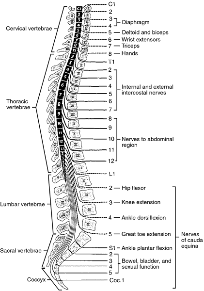

Spinal nerves emerging from the spinal cord through the intervertebral foramina with muscles or muscle movements listed for specific levels. From McQuillan et al., 2002.
splanchnic n's those of the blood vessels and viscera, especially the visceral branches of the thoracic, abdominal (lumbar), and pelvic parts of the sympathetic trunks. See Appendix 3-5.
sudomotor n's those that innervate the sweat glands.
sympathetic n's
1. see sympathetic trunk.
2. any nerve of the sympathetic nervous system.
trophic nerve one concerned with regulation of nutrition.
unmyelinated nerve one whose axons are not encased in a myelin sheath.
vasoconstrictor nerve one whose stimulation causes contraction of blood vessels.
vasodilator nerve one whose stimulation causes dilation of blood vessels.
vasomotor nerve one concerned in controlling the caliber of vessels, whether as a vasoconstrictor or vasodilator.
vasosensory nerve any nerve supplying sensory fibers to the vessels.
visceral nerve autonomic nerve.
Miller-Keane Encyclopedia and Dictionary of Medicine, Nursing, and Allied Health, Seventh Edition. © 2003 by Saunders, an imprint of Elsevier, Inc. All rights reserved.
nerve
(nerv), [TA]A whitish cordlike structure composed of one or more bundles (fascicles) of myelinated or unmyelinated nerve fibers, or more often mixtures of both, coursing outside the central nervous system, together with connective tissue within the fascicle and around the neurolemma of individual nerve fibers (endoneurium), around each fascicle (perineurium), and around the entire nerve and its nourishing blood vessels (epineurium), by which stimuli are transmitted from the central nervous system to a part of the body or the reverse. Nerve branches are given in the definition of the major nerve; many are also listed and defined under branch.
Synonym(s): nervus [TA]
[L. nervus]
Farlex Partner Medical Dictionary © Farlex 2012
nerve
(nûrv)n.
1. Any of the cordlike bundles of fibers made up of neurons through which sensory stimuli and motor impulses pass between the brain or other parts of the central nervous system and the eyes, glands, muscles, and other parts of the body. Nerves form a network of pathways for conducting information throughout the body.
2. The sensitive tissue in the pulp of a tooth.
3. nerves Nervous agitation caused by fear, anxiety, or stress: had a sudden attack of nerves.
4. A vein or rib in the wing of an insect.
The American Heritage® Medical Dictionary Copyright © 2007, 2004 by Houghton Mifflin Company. Published by Houghton Mifflin Company. All rights reserved.
nerve
Vox populi A popular adjective for cheekiness, brass, “balls,” as in she's got nerve. Cf Nerves.McGraw-Hill Concise Dictionary of Modern Medicine. © 2002 by The McGraw-Hill Companies, Inc.
nerve
(nĕrv) [TA]A whitish cordlike structure composed of one or more bundles (fascicles) of myelinated or unmyelinated nerve fibers, or more often mixtures of both, coursing outside of the central nervous system, together with connective tissue within the fascicle and around the neurolemma of individual nerve fibers (endoneurium), around each fascicle (perineurium), and around the entire nerve and its nourishing blood vessels (epineurium), by which stimuli are transmitted from the central nervous system to a part of the body or the reverse.
Synonym(s): nervus [TA] .
Synonym(s): nervus [TA] .
[L. nervus]
Medical Dictionary for the Health Professions and Nursing © Farlex 2012
nerve
A pinkish-white, cord-like structure consisting of bundles of long fibres (AXONS) of nerve cells and fine blood vessels held together by a connective tissue sheath. Individual fibres are usually insulated with a layer of white fatty material called myelin. The larger nerves contain both MOTOR and SENSORY fibres. Twelve pairs of nerves arise directly from the brain. These are called cranial nerves and carry impulses subserving smell, eye movement, vision, facial movement and sensation, all other sensation in the head, hearing, taste, movements of the soft palate, tongue and neck muscles, and control of the heartbeat and the secretion of stomach acid. 31 pairs of nerves emerge from the spinal cord. These control all the other muscles of the body and carry impulses for sensation from all parts of the body to the spinal cord and thence to the brain.Collins Dictionary of Medicine © Robert M. Youngson 2004, 2005
nerve
- (in animals) a bundle of nerve fibres, usually containing both afferent neurons (to the CENTRAL NERVOUS SYSTEM), and efferent neurones (away from CNS), together with associated connective tissue and blood vessels, situated in a common sheath of CONNECTIVE TISSUE and lying outside the CNS.
- (in plants) a characteristic leaf structure consisting of fine strands that are conductive and/or strengthening in function.
Collins Dictionary of Biology, 3rd ed. © W. G. Hale, V. A. Saunders, J. P. Margham 2005
Nerve
Fibers that carry sensory information, movement stimuli, or both from the brain and spinal cord to other parts of the body and back again. Some nerves, including the vagus nerve, innervate distantly separated parts of the body.
Mentioned in: Hiccups
Gale Encyclopedia of Medicine. Copyright 2008 The Gale Group, Inc. All rights reserved.
nerve
A whitish cord made up of myelinated or unmyelinated nerve fibres held together by connective tissue sheath in bundles and through which stimuli are transmitted from the central nervous system to the periphery or vice versa.
abducens nerve Sixth cranial nerve. It has its origin from the abducens nucleus at the lower border of the pons and at the lateral part of the pyramid of the medulla. It passes through the cavernous sinus and enters the orbit through the superior orbital fissure. It supplies motor innervation to the ipsilateral lateral rectus muscle. Additionally, interneurons leave the abducens nucleus and project to the contralateral medial rectus sub nucleus to allow conjugate gaze. A lesion in the nuclear region will cause gaze palsy, whereas an abducens nerve lesion will produce only an abduction deficit. See abducens nucleus; paralysis of the sixth nerve.
cranial nerve's Twelve pairs of nerves, one set on each side of the brain, that emerge, or enter the cranium. They carry sensory information from the sense organs, the muscles of the head, neck, shoulders, heart, viscera and vocal tract. The motor neurons with axons in the cranial nerves control pupil diameter, accommodation, movements of the eyes and eyelids, mastication, facial expression, head movements, as well as cardiorespiratory and digestive functions.
nerve fibre layer See retina.
fifth cranial nerve See trigeminal nerve.
fourth cranial nerve See trochlear nerve.
frontal nerve See ophthalmic nerve.
nerve impulse See action potential.
infratrochlear nerve See ophthalmic nerve.
lacrimal nerve See ophthalmic nerve.
long ciliary nerve One of a pair of nerves that comes off the nasociliary nerve and runs with the short ciliaries, pierces the sclera, travels in the suprachoroidal space and supplies sensory fibres to the iris, cornea, and ciliary muscle and sympathetic motor fibres to the dilator pupillae muscle (Fig. N2). See ophthalmic nerve; pupil light reflex.
nasociliary nerve See ophthalmic nerve.
oculomotor nerve Third cranial nerve. It is classified as a motor nerve. Its origin lies in the tegmentum of the midbrain. It passes through the cavernous sinus and just before it enters the orbit it divides into a small superior and a larger inferior division. Both divisions penetrate into the orbit through the superior orbital fissure. In the orbit the superior division passes inward above the optic nerve to supply the superior rectus and the levator palpebrae superioris muscles. The inferior division sends branches to the medial rectus, the inferior rectus and inferior oblique muscles, as well as providing parasympathetic fibres to the sphincter pupillae and ciliary muscles via a branch to the ciliary ganglion. See oculomotor nucleus; paralysis of the third nerve.
ophthalmic nerve This is the smallest of the three divisions of the trigeminal nerve, the other two being the maxillary and mandibular branches. It comes off the medial and upper part of the convex anterior border of the gasserian ganglion (trigeminal ganglion), passes through the cavernous sinus and just behind the superior orbital fissure it divides into three branches, the lacrimal, frontal and nasociliary, which pass through the fissure to enter the orbit. (1) The smallest of the three, the lacrimal nerve, supplies sensory fibres to the lacrimal gland, the skin of the upper eyelid and the conjunctiva. Just before reaching the gland the nerve communicates with the zygomaticotemporal nerve (itself a branch of the zygomatic nerve). This branch contains parasympathetic fibres from the facial nerve that pass to the lacrimal gland. (2) The frontal nerve, which is the largest of the three divisions, divides into the supratrochlear and supraorbital nerves. The supratrochlear further anastomoses with the infratrochlear nerve and supplies the lower part of the forehead, the upper eyelid and the conjunctiva. The infratrochlear supplies sensory fibres to the skin and conjunctiva round the inner angle of the eye, the root of the nose, the lacrimal sac and canaliculi and caruncle. The supraorbital nerve sends sensory fibres to the forehead, the upper eyelid and conjunctiva. (3) The nasociliary nerve gives origin to several nerves: the long ciliary nerves, the long or sensory root (ramus communicans) to the ciliary ganglion, the posterior ethmoidal nerve and the infratrochlear nerve (Fig. N2).
optic nerve Second cranial nerve. It forms a link in the visual pathway. It takes its origin at the retina and is made up of nearly 1.2 million fibres from the ganglion cells and some efferent fibres that end in the retina. The nerve runs backward from the eyeball and emerges from the orbit through the optic canal and then forms the optic chiasma. The total length of the optic nerve is 5 cm; the portion before the chiasma called intracranial being about 1 cm, the intracanalicular 6 mm, the intraorbital 3 cm and the intraocular 0.7 mm. The optic nerve is more often divided into only two portions: the intraocular (bulbar) portion and the orbital (retrobulbar) portion (Fig. N2). See optic atrophy; pupillary fibres; optic neuritis; anterior ischaemic optic neuropathy; papilloedema.
short ciliary nerve One of six to ten branches from the ciliary ganglion that enters the eye around the optic nerve, travels in the suprachoroidal space and innervates the ciliary muscle, the sphincter pupillae muscle and the cornea. See pupil light reflex.
sixth cranial nerve See abducens nerve.
supraorbital nerve; supratrochlear nerve See ophthalmic nerve.
third cranial nerve See oculomotor nerve.
trigeminal nerve Fifth cranial nerve. It is the largest of the cranial nerves. It originates above the middle of the lateral surface of the pons as two divisions, a larger sensory root and a motor root. The sensory root passes to the gasserian ganglion (trigeminal ganglion) and from that ganglion the three divisions of the fifth nerve are given off: the ophthalmic, maxillary and mandibular nerves. The fifth nerve is sensory to the face, the eyeball, the conjunctiva, the eyebrow, the teeth, the mucous membranes in the mouth and nose. The motor root of the nerve has no connection with the ganglion. It joins the mandibular nerve and is motor to the muscles of mastication.
trochlear nerve Fourth cranial nerve. It is the most slender of the cranial nerves but with the longest intracranial course (75 mm). It is the only motor nerve that originates from the dorsal surface of the brain between the midbrain and the cerebellum. It passes through the cavernous sinus and then enters the orbit through the superior orbital fissure and supplies motor fibres to the superior oblique muscle. See trochlear nucleus; paralysis of the fourth nerve.
zygomatic nerve A branch of the maxillary division of the trigeminal nerve, it enters the orbit by the inferior orbital fissure and soon divides into the zygomaticotemporal and zygomaticofacial branches. The former gives a twig to the lacrimal nerve and is thought to conduct autonomic fibres to the lacrimal gland and the latter supplies the skin over the zygomatic bone.
)
Fig. N2 Diagram of the ophthalmic division of the trigeminal nerve (fifth). This is a view of the right eye from above (c.b.z, communicating branch to the zygomatic nerve)
| Table N1 Cranial nerves | |||||
| nerve | type | function (sensory is in italic, the rest is motor) | |||
| I | olfactory | sensory | smell | ||
| II | optic | sensory | vision | ||
| III | oculomotor | mixed, primarily motor | movement of eye and eyelids, regulation of pupil size, accommodation, proprioception | ||
| IV | trochlear | mixed, primarily motor | eye movements, proprioception | ||
| V | trigeminal | mixed | chewing movements, sensations from head and face, proprioception | ||
| VI | abducens | mixed, primarily motor | abduction, proprioception | ||
| VII | facial | mixed | facial expression, secretion of saliva and tears, taste, proprioception | ||
| VIII | vestibulo-cochlear1. auditory (or cochlear) branch2. vestibular branch | sensory | hearingsense of balance | ||
| IX | glossopharyngeal | mixed | secretion of saliva, taste, control of blood pressure and respiration, proprioception | ||
| X | vagus | mixed | smooth muscle contraction and relaxation (e.g. heart) sensations from organs supplied, proprioception | ||
| XI | accessory | mixed, primarily motor | movements of head, swallowing movements and voice production, proprioception | ||
| XII | hypoglossal | mixed, primarily motor | tongue movements, proprioception | ||
Millodot: Dictionary of Optometry and Visual Science, 7th edition. © 2009 Butterworth-Heinemann
nerve
(nĕrv) [TA]A whitish cordlike structure composed of one or more bundles (fascicles) of myelinated or unmyelinated nerve fibers, or more often mixtures of both, coursing outside the central nervous system, together with connective tissue within the fascicle and around the neurolemma of individual nerve fibers (endoneurium), around each fascicle (perineurium), and around the entire nerve and its nourishing blood vessels (epineurium).
[L. nervus]
Medical Dictionary for the Dental Professions © Farlex 2012
Patient discussion about nerve
Q. why does ADHD make kind of an hype to children? is it a nerve defect?
A. it's a complex interaction among genetic and environmental factors causing a disorder in the central nervous system. a study showed a delay in development of certain brain structures n the frontal cortex and temporal lobe, which are believed to be responsible for the ability to control and focus thinking.
Q. What is ERD examination?My doctor want to find where is nerve is sprained. How this examonation will help? If the nerve is sprained by muscles or vertebrae what treat may be given by a doctor?
A. Sorry, but never heard of an examination called ERD, especially not for sprained muscle. Do you mean ERS?
Anyway, you may read more here:
www.nlm.nih.gov/medlineplus/sprainsandstrains.html
Anyway, you may read more here:
www.nlm.nih.gov/medlineplus/sprainsandstrains.html
Q. Nerves of pregnant woman can cause damage to the fetus?
A. Well, if by nerves you mean nervousness or stress, it hasn't been proven that stress during pregnancy can cause damage to the fetus. However, stress can damage the woman's health by lowering the body immunity and making it harder for the body to fight inflammation, infections, etc.
More discussions about nerveThis content is provided by iMedix and is subject to iMedix Terms. The Questions and Answers are not endorsed or recommended and are made available by patients, not doctors.