artery
[ahr´ter-e]a vessel through which the blood passes away from the heart to various parts of the body. The wall of an artery consists typically of an outer coat (tunica adventitia), a middle coat (tunica media), and an inner coat (tunica intima). 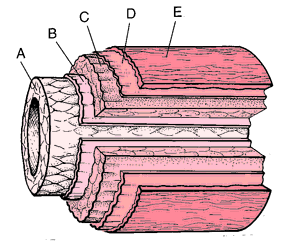 For names of specific arteries, see anatomic Table of Arteries in Appendices. See also Plate 8.

Representation of arterial coats: A, tunica intima; B, internal elastic lamina; C, tunica media; D, external elastic lamina; E, tunica externa. From Dorland's, 2000.
end artery one that undergoes progressive branching without development of channels connecting with other arteries.
nutrient artery any artery that supplies the marrow, or medulla, of a long bone.
Miller-Keane Encyclopedia and Dictionary of Medicine, Nursing, and Allied Health, Seventh Edition. © 2003 by Saunders, an imprint of Elsevier, Inc. All rights reserved.
ar·ter·y (a),
(ar'ter-ē), [TA]A relatively thick-walled, muscular, pulsating blood vessel conveying blood away from the heart. With the exception of the pulmonary and umbilical arteries, the arteries contain red or oxygenated blood. At the major arteries, the arterial branches are listed separately following the designation branches.
Synonym(s): arteria [TA]
[L. arteria, fr. G. artēria]
Farlex Partner Medical Dictionary © Farlex 2012
artery
(är′tə-rē)n. pl. arter·ies
1. Anatomy Any of the muscular elastic tubes that form a branching system and that carry blood away from the heart to the cells, tissues, and organs of the body.
2. A major route of transportation into which local routes flow: Traffic was heavy on the central artery.
The American Heritage® Medical Dictionary Copyright © 2007, 2004 by Houghton Mifflin Company. Published by Houghton Mifflin Company. All rights reserved.
ar·te·ry
(ahrtĕr-ē ) [TA]A generally (with the exception of the coronary artery), muscular blood vessel conveying blood away from the heart to the tissues. With the exception of the pulmonary and umbilical arteries, the arteries convey red or oxygenated blood.
Synonym(s): arteria [TA] .
Synonym(s): arteria [TA] .
[L. arteria, fr. G. artēria]
Medical Dictionary for the Health Professions and Nursing © Farlex 2012
artery
An elastic, muscular-walled tube carrying blood at high pressure from the heart to any part of the body. From the Greek arteria , an air duct. It was once believed that, in life, arteries contained air.Collins Dictionary of Medicine © Robert M. Youngson 2004, 2005
)
Fig. 49 Artery . Transverse sections.
artery
any of the blood vessels with thick, elasticated muscular walls that carry blood to the tissues from the heart, forming part of the BLOOD CIRCULATORY SYSTEM.Arteries usually carry oxygenated blood but the pulmonary artery carries deoxygenated blood from the right ventricle to the lungs. Arteries become less massive as the vessels approach their target organs, eventually becoming reduced to ARTERIOLES. See VEIN.
Collins Dictionary of Biology, 3rd ed. © W. G. Hale, V. A. Saunders, J. P. Margham 2005
Artery
One of several tubular branches of muscular and elastic walled vessels that carry blood from the heart through the body.
Gale Encyclopedia of Medicine. Copyright 2008 The Gale Group, Inc. All rights reserved.
artery
A tubular, elastic vessel which carries blood away from the heart. Its walls are thicker than those of veins in order to withstand the greater pressure of blood on the arterial side of the circulation.
anterior ciliary artery's See ciliary artery.
central retinal artery A branch of the ophthalmic artery entering the optic nerve some 6-12 mm from the eyeball. It enters the eye through the optic disc and divides into superior and inferior branches. Both these branches subdivide into nasal and temporal branches which course in the nerve fibre layer, supplying the capillaries feeding the bipolar and the ganglion cell layers of the retina (except for the foveola). The outer third of the retina containing the photoreceptors is supplied by the choriocapillaris. See temporal arteritis; retinal arterial occlusion; cherry-red spot; central retinal vein.
carotid artery See amaurosis fugax; internal carotid artery.
ciliary artery's Branches of the ophthalmic artery that supply the whole of the uveal tract, the sclera and the edge of the cornea with its neighbouring conjunctiva. The ophthalmic artery gives rise to one lateral and one medial posterior ciliary artery. The latter divides into the short and the long posterior ciliary arteries.The short posterior ciliary arteries are some 10-20 branches that pierce the eyeball in an irregular ring around the optic nerve to supply the posterior choroid, the optic disc, the circle of Zinn and the cilioretinal arteries. The long posterior ciliary arteries are two branches, which pierce the sclera between 3 and 4 mm from the optic nerve on either side and course in the suprachoroidal space (Fig. A19). They form, with the anterior ciliary arteries, the major arterial (or iridic) circle of the iris, which supplies the ciliary body, the anterior choroid and the iris. The anterior ciliary arteries are derived from the arteries to the four recti muscles and they anasto-mose in the ciliary muscle with the long posterior ciliary arteries to form the major arterial circle of the iris. They also give branches that supply the episclera (episcleral arteries), sclera, limbus and conjunctiva (anterior and posterior conjunctival arteries). See anastomosis; major arterial circle of the iris.
cilioretinal artery A small artery running from the temporal side of the optic disc to the macular area. It originates from the circle of Zinn and supplies the retina between the macula and the disc. This artery is present in only about a fifth, or less, of human eyes. If a patient possesses this artery central vision will be spared in case of occlusion of the central retinal artery. In some other eyes the cilioretinal artery supplies some other region of the retina. See circle of Zinn.
conjunctival artery's See ciliary arteries.
copper wire artery See arteriosclerosis.
hyaloid artery An artery that is present during the embryological period. It arises from the ophthalmic artery, runs forward from the optic disc to the lens where it spreads over the posterior lenticular surface as a capillary net which in turn anastomoses with a capillary net located on the anterior lens surface. Thus the lens becomes enveloped by an anastomosing vascular network called the tunica vasculosa lentis. The hyaloid artery also gives rise to a large number of branches, the vasa hyaloidea propria, which at times almost fills the vitreous cavity. The hyaloid artery degenerates by the eighth month of gestation to become the central retinal artery. See hyaloid canal; optic fissure; hyaloid remnant.
infraorbital artery A terminal branch of the internal maxillary artery which enters the orbit through the inferior orbital fissure and appears on the face via the infraorbital canal. It supplies the inferior rectus and inferior oblique muscles, the lacrimal sac, the lower eyelid, the upper teeth and lip.
internal carotid artery A branch of the common carotid artery. The internal carotid artery gives rise to many branches and in particular the ophthalmic artery after it passes through the cavernous sinus. It terminates in the anterior and middle cerebral arteries (Fig. A19). See amaurosis fugax; circle of Willis; Hollenhorst's plaques.
lacrimal artery It arises from the ophthalmic artery to the outer side of the optic nerve. It supplies the lacrimal gland, the conjunctiva and eyelids, giving origin to the lateral palpebral arteries (Fig. A19).
ophthalmic artery Vessel arising from the internal carotid artery and which enters the orbit through the optic canal. It gives rise to numerous branches: (1) Central retinal artery. (2) Posterior ciliary arteries. (3) Lacrimal artery (and lateral palpebral and zygomatic branches). (4) Muscular branches. (5) Supraorbital artery. (6) Anterior and posterior ethmoidal arteries. (7) Recurrent meningeal artery. (8) Supratrochlear artery. (9) Medial palpebral arteries. (10) Dorsal nasal artery.Thus, the ophthalmic artery supplies all the tunics of the eyeball, most of the structures in the orbit, the lacrimal sac, the paranasal sinuses, and the nose (Fig. A19).
silver wire artery See arteriosclerosis.
supraorbital artery Branch of the ophthalmic artery which supplies the upper eyelid, the scalp and also sends branches to the levator palpebrae superioris muscle and the periorbita (Fig. A19).
)
Fig. A19 The ophthalmic artery and its branches
Millodot: Dictionary of Optometry and Visual Science, 7th edition. © 2009 Butterworth-Heinemann
ar·te·ry
(ahrtĕr-ē ) [TA]A relatively thick-walled, muscular, pulsating blood vessel conveying blood away from the heart.
Synonym(s): arteria [TA] .
Synonym(s): arteria [TA] .
[L. arteria, fr. G. artēria]
Medical Dictionary for the Dental Professions © Farlex 2012
Patient discussion about artery
Q. my mother have stem replacement for a coronary artery oclusion is already 2 years she physically deteriorating since surgery why???? please help she does not have energy
A. I agree with Dagmar. It can be most likely caused by another occlusion or re-occlusion inside the heart blood vessels. Since that is a life-threatening case, I strongly suggest you to bring your mother into a hospital (for complete check up), or just call your cardiologist to have first treatment.
Meanwhile, that will be better if you have emergency oxygen (just in case you'll need it) with you.
Meanwhile, that will be better if you have emergency oxygen (just in case you'll need it) with you.
Q. how many 1. calories 2. good vs bad fat 3. protein does 1 cup of whole milk have compared to 1 cup of almonds?
A. Each almond has 7 calories. A cup of almonds has 680 calories, Total Fat: 60g, out of which 3.9g are Saturated Fat (=bad fat), Carbs: 24g, Protein: 24g.
1 cup of 2% milk has 130 calories, Total Fat: 5g, out of which 3g are Saturated Fat (=bad fat), Carbs: 13g, Protein: 8g.
Here is the nutrition value of different kinds of milk as well:
http://www.cassclay.com/milk_nut.html
More discussions about artery1 cup of 2% milk has 130 calories, Total Fat: 5g, out of which 3g are Saturated Fat (=bad fat), Carbs: 13g, Protein: 8g.
Here is the nutrition value of different kinds of milk as well:
http://www.cassclay.com/milk_nut.html
This content is provided by iMedix and is subject to iMedix Terms. The Questions and Answers are not endorsed or recommended and are made available by patients, not doctors.