muscle
[mus´'l]Muscle fibers range in length from a few hundred thousandths of a centimeter to several centimeters. They also vary in shape, and in color from white to deep red. Each fiber receives its own nerve impulses, so that fine and varied motions are possible. Each has its small stored supply of glycogen, which it uses as fuel for energy. Muscles, especially the heart, also use free fatty acids as fuel. At the signal of an impulse traveling down the nerve, the muscle fiber changes chemical energy into mechanical energy, and the result is muscle contraction.
Some muscles are attached to bones by tendons. Others are attached to other muscles, or to skin (producing the smile, the wink, and other facial expressions, for example). All or part of the walls of hollow internal organs, such as the heart, stomach, intestines, and blood vessels, are composed of muscles. The last stages of swallowing and of peristalsis are actually series of contractions by the muscles in the walls of the organs involved.
Involuntary muscles are those not under the control of the conscious part of the brain; they respond to the nerve impulses of the autonomic nervous system. They include the countless short-fibered, or smooth, muscles of the internal organs and power the digestive tract, the pupils of the eyes, and all other involuntary mechanisms.
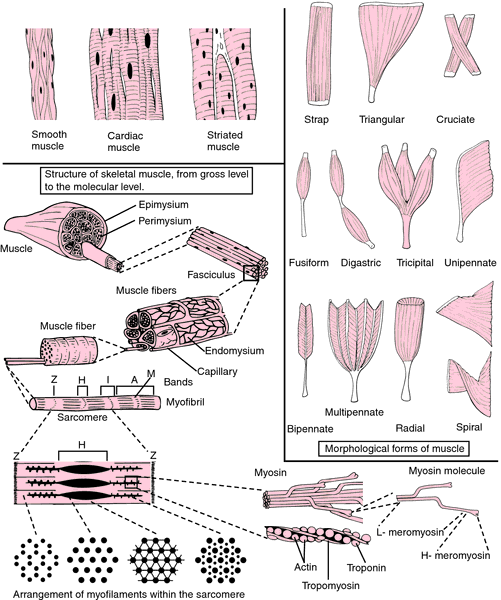
Voluntary muscles are those controlled by the conscious part of the brain, and are striated. These are the skeletal muscles that enable the body to move, and there are more than 600 of them in the human body. Their fibers are grouped together in sheaths of muscle cells. Groups of fibers are bundled together into fascicles, surrounded by a tough sheet of connective tissue to form a muscle group such as the biceps. Unlike the involuntary muscles, which can remain in a state of contraction for long periods without tiring and are capable of sustained rhythmic contractions, the voluntary muscles are readily subject to fatigue.
Cardiac muscles (the muscles of the heart) are the third kind; they are involuntary and consist of striated fibers different from those of voluntary muscle. The contraction and relaxation of cardiac muscle continues at a rhythmic pace until death unless the muscle is injured in some way.

Muscles enable the body to perform different types of movement. Those that bend a limb at a joint, raising a thigh or bending an elbow, are called flexors. Those that straighten a limb are called extensors. Others, the abductors, make possible movement away from the midline of the body, whereas the adductors permit movement toward the midline. Muscles always act in opposing groups. In bending an elbow or flexing a muscle, for example, the biceps (flexor) contracts and the triceps (extensor) relaxes. The reverse happens in straightening the elbow.
A muscle that has contracted many times, and has exhausted its stores of glycogen and other substances, and accumulated too much lactic acid, becomes unable to contract further and suffers from fatigue. In prolonged exhausting work, fat in the muscles can also be used for energy, and as a consequence the muscles become leaner.
mus·cle
(mŭs'ĕl), [TA]muscle
(mŭs′əl)MUSCLE
Abbreviation for:Multi Station Clinical Examination (OSCE)
mus·cle
(mŭs'ĕl) [TA]Synonym(s): musculus.
muscle
A tissue consisting of large numbers of parallel elongated cells with the power of shortening and thickening so as to approximate their ends and effect movement. Up to 50% of the body weight consists of muscle, most being attached to bone in such a way that muscle contraction causes joints to bend (flex) or straighten (extend). Muscle fibres convert chemical energy into mechanical energy. There are three kinds of muscle—striped (striated) or voluntary muscle; smooth or involuntary muscle occurring in the walls of arteries, the intestines and the urinary tract; and heart muscle (MYOCARDIUM), a network (syncytium) of muscle fibres that contract regularly and automatically without external stimulus.)
muscle
the fleshy part of any animal that consists of tissue made up of highly contractile cells which serve to move parts of the body relative to each other.A muscle is composed of many fibres or muscle cells. In STRIATED MUSCLE, each cell contains a bundle of MYOFIBRILS each exhibiting a banding pattern and being made up of a number of SARCOMERES arranged end to end. The sarcomere is the unit of contraction and the banding visible over its surface results from the longitudal filaments which make up the myofibril being of two types, thick (dark) and thin (light). These filaments overlap as shown in Fig. 225. The thick filaments are composed of the protein MYOSIN and the thin filaments of ACTIN. H.E. Huxley and K. Harrison found that on contraction, the light zones (I-BANDS) were comparatively narrow; on relaxation of the muscle the I-bands were broad. Where very strong contraction takes place the H-zone disappears and the thin filaments overlap.
Huxley and Harrison proposed the sliding filament hypothesis, to account for their observations. Bridges occur between thick and thin filaments and in contraction the bridges pull thin filaments past the thick ones using a ratchet mechanism. Some filaments are retained in this ‘pulled past’ position whilst others detach then reattach and repeat the ‘pulling past’ action. ACTOMYSIN is formed at the point of contact of bridge and thin filament. For each bridge to go through its cycle of attachment, contraction and reattachment, the splitting of one molecule of ATP is required, the cycles occurring between 50 and 100 times per second. The supply of ATP comes from MITOCHONDRIA between the fibrils. Calcium ions are released from vesicles in the sarcoplasmic reticulum, by the ACTION POTENTIAL passing along the surface of the fibre and these split the ATP. Troponin activated by the calcium displaces tropomyosin, which prevents myosin bridges from binding with actin fibrils. Once binding takes place this activates ATPase and on hydrolysis of ATP the bridge goes through its cycle of movement.
muscle
abducens muscle See lateral rectus muscle.
adducens muscle See medial rectus muscle.
agonistic muscle A muscle that performs the desired movement, or does the opposite to an antagonistic muscle. Example: the left lateral rectus is the agonistic muscle when the left eye turns to the left. See antagonistic muscle.
antagonistic muscle A muscle that opposes the action of another. Example: the right superior rectus muscle is the contralateral antagonist of the left superior oblique. See agonistic muscle; synergistic muscles.
Brücke's muscle See ciliary muscle.
ciliary muscle The smooth (unstriated and involuntary) muscle of the ciliary body. In a meridional section of the eye it has the form of a right-angled triangle, the right angle being internal and facing the ciliary processes. The posterior angle is acute and points to the choroid, the hypotenuse runs parallel with the sclera. Some of its fibres have their origin in the scleral spur at the angle of the anterior chamber, while other fibres take origin in the trabecular meshwork. The fibres radiate backward in three directions: (1) Fibres coursing meridionally or longitudinally more or less parallel to the sclera and can be traced posteriorly into the suprachoroid to the equator or even beyond. They end usually in branched stellate figures known as muscle stars with three or more rays to each. These fibres represent Brücke's muscle. (2) Other fibres course radially. These fibres lie deep in the longitudinal fibres from which they are distinguished by the reticular character of their stroma but are often very difficult to separate from the circular fibres. (3) The circular fibres (Müller's muscle) occupy the anterior and inner portion of the ciliary body and run parallel to the limbus. As a whole, these fibres form a ring.Innervation to the ciliary muscle (mainly parasympathetic fibres derived from the oculomotor nerve) is provided through the short ciliary nerves and stimulation causes a contraction of the muscle. However, a small amount of sympathetic supply is also believed to act and relax the muscle. Blood supply to the ciliary muscle is provided by the anterior and long posterior ciliary arteries. Contraction of the ciliary muscle causes a reduction in its length thus causing the whole muscle to move forward and inward. Consequently the zonule of Zinn, which suspends the lens, relaxes. This leads to a decrease in the tension in the capsule of the lens allowing it to become more convex and thereby providing accommodation. Syn. Bowman's muscle. See mechanism of accommodation; adrenergic receptors; ciliary body; scleral spur; Helmholtz's of accommodation theory; zonule of Zinn.
muscle cone A structure formed by the sheath of the four recti muscles as they pass forward from their common origin at the apex of the orbit in the fibrous ring called the annulus of Zinn (and around the optic nerve) to be inserted into the sclera around the eyeball. Some authors consider the muscle cone to include the superior oblique muscle. See annulus of Zinn.
dilator pupillae muscle Smooth (unstriated and involuntary) muscle whose fibres constitute the posterior membrane of the iris. This muscle extends from the ciliary body close to the margin of the iris where it fuses with the sphincter pupillae muscle. Contraction of the dilator pupillae muscle draws the pupillary margin towards the ciliary body and therefore dilates the pupil. This muscle is supplied by the sympathetic fibres in the long ciliary nerves and by a few parasympathetic fibres. See adrenergic receptors; sphincter pupillae muscle; mydriatic.
elevator muscle's See inferior oblique muscle; superior rectus muscle.
external rectus muscle See lateral rectus muscle.
extraocular muscle's The striated (voluntary) muscles that control the movements of the eyes. There are six such muscles: four recti muscles (lateral rectus, medial rectus, superior rectus and inferior rectus) which move the eye more or less around the transverse and vertical axes, and two oblique muscles (inferior oblique and superior oblique) which move the eyes obliquely. The muscles are composed of striated fibres of varying length, mostly running parallel to the direction of the muscle and united by fibrous connective tissue. They have a greater ratio of nerve fibres to muscle fibres than other striated muscles of the body. The fibre thickness varies from 3 to 50 μm, although functionally there seem to be two main types of fibres, the fast and the slow fibres. The former are the thickest and probably responsible for the fast movements of the eyes (saccades) and the latter consist of thin fibres. The tendons (bands of connective tissue) at one end of each extraocular muscle are attached to bones. This is the origin of the muscle. At the other end of the muscle the tendon is attached to the eye and this area is called the insertion. The substance proper of the muscle is called the belly. Contraction of a muscle occurs in the direction of its constituent fibres and causes a shortening of the muscle. Consequently the eye turns in a given direction depending upon which extraocular muscle is contracting. Contraction results from nervous impulses arriving at the motor end-plate (the junction between an axon and a striated muscle fibre) of the muscle through one of the ocular motor nerves. This causes a neurotransmitter substance to be discharged in the microscopic gap between the end-plate and a muscle fibre. These muscles also possess specialized receptors called muscle spindles, which are small groups of muscle fibres that are provided with both a sensory and a motor nerve supply. There are between 12 and 50 in each muscle. The muscle spindles provide a constant and continuous monitoring of the degree of tension of the muscle itself. Syn. extrinsic muscles; oculorotary muscles. See cholinergic; felderstruktur fibres; motor unit; strabismus surgery; motility test; three-step test.
extrinsic muscle's See extraocular muscles.
eyelid retractor muscle's See levator palpebrae superioris muscle; Müller's palpebral muscles.
Horner's muscle A thin layer of fibres that originates behind the lacrimal sac from the upper part of the posterior lacrimal crest (a ridge on the lacrimal bone which borders the fossa for the lacrimal sac). The muscle passes outward and forward and divides into two slips surrounding the canaliculi. It then becomes continuous with the pretarsal portions of the orbicularis muscle of the upper and lower lids and with the muscle of Riolan. Horner's muscle may be involved in tear drainage through action on the lacrimal sac. Syn. pars lacrimalis muscle; tensor tarsi muscle. See muscle of Riolan.
inferior oblique muscle (IO) One of the extraocular muscles, it takes its origin at the antero-medial corner of the floor of the orbit. It passes underneath the inferior rectus in a backward direction (making an angle of about 50º with the sagittal plane of the eye), then under the lateral rectus to be inserted by the shortest tendon of all extraocular muscles on the posterior, temporal portion of the eyeball, for the most part below the horizontal meridian, some 5 mm away from the optic nerve. It is innervated by the oculomotor nerve and it extorts (main action), elevates and abducts the eyeball when the eye is in the primary position. Combined with the action of the superior rectus muscle, it directs the eye upward (Fig. M15). See Bielschowsky's head tilt test; three-step test.
inferior rectus muscle (IR) This is the shortest of the four recti muscles. It arises from the lower part of the annulus of Zinn, runs forward, downward and outward (making an angle of about 23º with the sagittal plane) and inserts into the inferior portion of the sclera about 6.5 mm from the corneal limbus. It is innervated by the inferior division of the oculomotor nerve and it depresses (main action), adducts and extorts the eyeball when the eye is in the primary position (Fig. M15). See annulus of Zinn; Müller's palpebral muscles; Bielschowsky's head tilt test.
inferior tarsal muscle See Müller's palpebral muscles.
internal rectus muscle See medial rectus muscle.
intraocular muscle's The smooth (unstriated and involuntary) muscles found within the eye. They are the ciliary, the dilator pupillae and the sphincter pupillae muscles. Syn. intrinsic muscles. See cholinergic.
lateral rectus muscle (LR) One of the extraocular muscles, it arises from both the lower and upper parts of the annulus of Zinn which bridge the superior orbital fissure. The muscle passes forward along the lateral wall of the orbit, crosses the tendon of the inferior oblique muscle and inserts into the sclera about 6.9 mm from the corneal limbus. It is innervated by the abducens nerve and it abducts the eyeball when the eye is in the primary position (Fig. M15). Syn. external rectus muscle; abducens muscle. See annulus of Zinn; check ligament.
levator palpebrae superioris muscle Striated muscle that arises from the under surface of the lesser wing of the sphenoid bone above and in front of the optic canal. It passes forward below the roof of the orbit and above the superior rectus muscle and terminates in a tendinous expansion or aponeurosis (also called levator aponeurosis), which spreads out in a fan-shaped manner so as to occupy the whole breadth of the orbit and thus gives the whole muscle the form of an isosceles triangle. From the inferior surface of the aponeurosis arises a thin sheet of smooth muscle fibres called Müller's palpebral muscle (or superior tarsal muscle) which inserts into the posterior margin of the superior tarsal plate and into the superior fornix of the conjunctiva. These smooth muscle fibres are innervated by sympathetic nerves from the superior cervical sympathetic ganglion and assist in elevating the upper eyelid. The fibres of the aponeurosis are attached to the anterior margin of the superior tarsal plate while some fuse with bundles of the orbicularis oculi muscle to attach to the skin. These latter sets of fibres produce the horizontal skin crease of the upper eyelid. The striated levator aponeurosis is innervated by the superior division of the oculomotor nerve and elevates the upper eyelid. Its antagonist is the orbicularis muscle. See orbicularis muscle.
medial rectus muscle (MR) One of the extraocular muscles, it arises from the medial part of the annulus of Zinn. It passes forward along the medial wall of the orbit and is inserted into the sclera about 5.5 mm from the corneal limbus. It is innervated by the inferior division of the oculomotor nerve and it adducts the eyeball when the eye is in the primary position (Fig. M15). Syn. internal rectus muscle; adducens muscle. See annulus of Zinn; check ligament.
Müller's muscle See ciliary muscle.
Müller's palpebral muscle's Smooth muscles of the eyelids. The superior one (also called superior tarsal muscle) originates from the under surface of the levator palpebrae superioris muscle and passes below to insert into the upper margin of the tarsal plate of the upper eyelid. The inferior one (also called inferior tarsal muscle) originates from the muscular fascia covering the inferior rectus muscle. It extends upward and inserts into the bulbar conjunctiva and the lower margin of the tarsal plate of the lower eyelid. Müller's palpebral muscles are innervated by sympathetic fibres and help in lifting the upper eyelid and depressing the lower eyelid. They are sometimes referred to as the eyelid retractors.
oculorotary muscle's See extraocular muscles.
orbicularis muscle A thin oval sheet of striated muscle that surrounds the palpebral fissure, covers the eyelids and spreads out for some distance onto the temple, forehead and cheek. It consists of three portions: (1) The marginal or ciliary portion (muscle of Riolan). (2) The palpebral portion (also called the pars palpebralis muscle) which is the essential part of the muscle and is confined to the lids and may itself be divided into pretarsal portion whose fibres lie in front of the tarsal plates, and the preseptal portion whose fibres extend from the tarsal plates to the orbital margin. The palpebral portion is used in closing the eye without effort or in reflex blinking. (3) The orbital portion (also called the pars orbitalis muscle) which is found in the eyebrow, the temple, the forehead and the cheek. This portion of the muscle is used to close the eye tightly and the skin of the forehead, temple and cheek is drawn towards the inner side of the orbit. The orbicularis muscle is innervated by the facial nerve. Syn. sphincter oculi muscle. See ectropion; Horner's muscle; levator palpebrae superiotis muscle; muscle of Riolan; myokymia.
pars ciliaris muscle See muscle of Riolan.
pars lacrimalis muscle See Horner's muscle.
pars orbitalis muscle See orbicularis muscle.
pars palpebralis muscle See orbicularis muscle.
pupillary muscle's The dilator pupillae and the sphincter pupillae muscles.
muscle of Riolan The ciliary portion of the orbic-ularis muscle, it consists of very fine striated muscle fibres which lie in the dense tissue of the eyelids near their margin. It is continuous with Horner's muscle and encircles the eyelid margins mainly between the tarsal glands and the eyelash follicles. Its action is to bring the eyelid margins together when the eyes are closed. Syn. pars ciliaris muscle. See Horner's muscle; orbicularis muscle.
sphincter oculi muscle See orbicularis muscle.
sphincter pupillae muscle Smooth, circular muscle about 1 mm broad, forming a ring all round the pupillary margin near the posterior surface of the iris. It is innervated by parasympathetic fibres of the oculomotor nerve that synapse in the ciliary ganglion and by a few sympathetic fibres. Its contraction produces a reduction in the diameter of the pupil. See miotics; dilator pupillae muscle; pupil light reflex.
muscle spindle See extraocular muscles.
superior oblique muscle (SO) This is the longest and thinnest of the extraocular muscles. It arises above and medial to the optic foramen on the small wing of the sphenoid bone. It passes forward between the roof and medial wall of the orbit to the trochlea (which is in the form of a pulley made of fibrocartilage) located at the front of the orbit where it loops over and turns sharply backward, downward and outward (making an angle of about 55º with the sagittal plane), passes under the superior rectus and inserts into the sclera just behind the equator on the superior temporal portion of the eyeball. It is innervated by the trochlear nerve and it intorts (main action), depresses, and also abducts the eyeball when the eye is in the primary position (Fig. M15). See trochlear fossa; Bielschowsky's head tilt test; three-step test.
superior rectus muscle (SR) One of the extraocular muscles, it arises from the upper part of the annulus of Zinn. It passes forward and outward (making an angle of about 23º with the sagittal plane) and inserts into the sclera about 7.7 mm from the corneal limbus. It is innervated by the superior division of the oculomotor nerve and elevates (main action), adducts, and also intorts the eyeball when the eye is in the primary position (Fig. M15). See annulus of Zinn; Bielschowsky's head tilt test.
synergistic muscle's Muscles having a similar and mutually helpful action as, for example, the inferior rectus and superior oblique muscles in depressing the eyeball. See Table M5.
superior tarsal muscle See Müller's palpebral muscles.
tarsal muscle's See Müller's palpebral muscles.
tensor tarsi muscle See Horner's muscle.
yoke muscle's Muscles of the two eyes which simultaneously contract to turn the eyes in a given direction. Example: the medial rectus of the right eye and the lateral rectus of the left eye when turning the eyes to the left. See Hering's law of equal innervation; motility test; version.
)
| Table M5 Agonistic, antagonistic and synergistic extraocular muscles | ||||||
| agonist | ipsilateral antagonist | ipsilateral synergist(s) | contralateral synergist | |||
| lateral rectus | medial rectus | superior oblique | medial rectus | |||
| inferior oblique | ||||||
| medial rectus | lateral rectus | superior rectus | lateral rectus | |||
| inferior rectus | ||||||
| superior rectus | inferior rectus | inferior oblique | inferior oblique | |||
| inferior rectus | superior rectus | superior oblique | superior oblique | |||
| superior oblique | inferior oblique | superior rectus | inferior rectus | |||
| inferior oblique | superior oblique | inferior rectus | superior rectus | |||
| Table M6 Innervation and action of the six extraocular muscles | ||||
| muscle | innervation | action in the primary position | ||
| medial rectus | oculomotor (III) | adduction | ||
| lateral rectus | abducens (VI) | abduction | ||
| inferior rectus | oculomotor (III) | depression* | ||
| adduction | ||||
| extorsion | ||||
| superior rectus | oculomotor (III) | elevation | ||
| adduction | ||||
| intorsion | ||||
| inferior oblique | oculomotor (III) | extorsion | ||
| elevation | ||||
| abduction | ||||
| superior oblique | trochlear (IV) | intorsion | ||
| depression | ||||
| abduction | ||||
| *Bold characters indicate main action. | ||||
| Table M7 Intraocular muscles of the eyeball (unstriated muscles) | ||
| name of muscle | nerve supply | action |
| sphincter pupillae | parasympathetic via oculomotor nerve | constricts pupil |
| dilator pupillae | sympathetic via trigeminal nerve | dilates pupil |
| ciliary | parasympathetic via oculomotor nerve | controls shape of lens in accommodation |
| Table M8 Yoke muscles | ||
| right eye | left eye | version* |
| lateral rectus | medial rectus | to the right |
| medial rectus | lateral rectus | to the left |
| superior rectus | inferior oblique | up and to the right |
| inferior rectus | superior oblique | down and to the right |
| superior oblique | inferior rectus | down and to the left |
| inferior oblique | superior rectus | up and to the left |
| *The directions refer to those of the patient. | ||
| Table M9 Dimensions of the four recti muscles | ||||||
| muscle length (mm) | insertion distance from limbus (mm) | tendon length (mm) | ||||
| lateral rectus | 48 | 6.9 | 8.8 | |||
| medial rectus | 40 | 5.5 | 3.7 | |||
| inferior rectus | 40 | 6.5 | 5.5 | |||
| superior rectus | 42 | 7.7 | 5.8 | |||
mus·cle
(mŭs'ĕl) [TA]Synonym(s): musculus [TA] .
Patient discussion about muscle
Q. What are muscle cramps caused from? I am a 30 year old woman and am pregnant. I keep on getting a muscle cramps on the back on my lower leg. It really hurts! What is causing it and how can I prevent it?
No one knows for certain what causes leg cramps in pregnancy, though there are some theories: Deficiencies in salt, calcium, magnesium and vitamin C or changes in blood circulation.
To prevent it make sure to stretch your muscles before bed and if you do get a cramp, immediately stretch your calf muscles: Straighten your leg, heel first, and gently flex your toes back toward your shins. It might hurt at first, but it will ease the spasm and the pain will gradually go away.
Q. Why do my muscles sometimes burn when I'm exercising? I do exercise twice a day. Why do my muscles sometimes burn when I'm exercising?
Muscle cells take up glucose (muscle glycogen) and convert them into lactic acid, which the mitrochondria in the cells then use for energy. The old theory was that lactic acid was a waste product that hindered performance. New scholarship on this actually shows that lactic acid is a SOURCE of fuel, not a "dead end as far as energy production is concerned."
Much of this new thinking has come from research performed by Dr. George Brooks at the University of California - Berkely. You can read more here: http://berkeley.edu/news/media/releases/2006/04/19_lactate.shtml
Researchers also now believe that muscle acidosis (that burning sensation during exercise)is not caused by increases in lactate within the muscle, but rather by a completely separate reaction when ATP is h
Q. What can I do to build muscle and develop immunity? I'm Mickey, 21. My height is 5’5” and I weigh 176 lbs. I love out door games especially soccer. I have poor immunity that I get sick very often. What can I do to build muscle and develop immunity?