laparoscopy
[lap″ah-ros´kah-pe]examination by means of the laparoscope; called also celioscopy. adj., adj laparoscop´ic.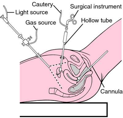

Laparoscopy. From Frazier et al., 2000.
Miller-Keane Encyclopedia and Dictionary of Medicine, Nursing, and Allied Health, Seventh Edition. © 2003 by Saunders, an imprint of Elsevier, Inc. All rights reserved.
per·i·to·ne·os·co·py
(per'i-tō-nē-os'kŏ-pē),Examination of the contents of the peritoneum with a peritoneoscope passed through the abdominal wall. See: laparoscopy.
Synonym(s): celioscopy, ventroscopy
Farlex Partner Medical Dictionary © Farlex 2012
celioscopy
(sē′lē-ŏs′kə-pē)n.
See peritoneoscopy.
The American Heritage® Medical Dictionary Copyright © 2007, 2004 by Houghton Mifflin Company. Published by Houghton Mifflin Company. All rights reserved.
ce·li·os·co·py
(sē'lē-os'kŏ-pē) Synonym(s): peritoneoscopy, coelioscopy.
[celio- + G. skopeō, to view]
Medical Dictionary for the Health Professions and Nursing © Farlex 2012