Conjunctivitis
Definition
Conjuctivitis is an inflammation or redness of the lining of the white part of the eye and the underside of the eyelid (conjunctiva) that can be caused by infection, allergic reaction, or physical agents like infrared or ultraviolet light.
Description
Conjunctivitis is the inflammation of the conjunctiva, a thin, delicate membrane that covers the eyeball and lines the eyelid. Conjunctivitis is an extremely common eye problem because the conjunctiva is continually exposed to microorganisms and environmental agents that can cause infections or allergic reactions. Conjunctivitis can be acute or chronic depending upon how long the condition lasts, the severity of symptoms, and the type of organism or agent involved. It can also affect one or both eyes and, if caused by infection, can be very easily transmitted to others during close physical contact, particularly among children in a daycare center. Other names for conjunctivitis include pink eye and red eye.
Causes and symptoms
Conjunctivitis may be caused by a viral infection, such as a cold, acute respiratory infection, or disease such as measles, herpes simplex, or herpes zoster. Symptoms include mild to severe discomfort in one or both eyes, redness, swelling of the eyelids, and watery, yellow, or green discharge. Symptoms may last anywhere from several days to two weeks. Infection with an adenovirus, however, may also cause a significant amount of pus-like discharge and a scratchy, foreign body-type of sensation in the eye. This may also be accompanied by swelling and tenderness of the lymph nodes near the ear.
Bacterial conjunctivitis can occur in adults and children and is caused by organisms such as Staphylococcus, Streptococcus, and Hemophilus. Symptoms of bacterial conjunctivitis include a pus-like discharge and crusty eyelids after awakening. Redness of the conjunctiva can be mild to severe and may be accompanied by swelling. Persons with symptoms of conjunctivitis who are sexually active may possibly be infected with the bacteria that cause either gonorrhea or chlamydia. There may be large amounts of pus-like discharge, and symptoms may include intolerance to light (photophobia), watery mucous discharge, and tenderness in the lymph nodes near the ear that may persist for up to three months.
Conjunctivitis may also be caused by environmental hazards, such as wind, smoke, dust, and allergic reactions caused by pollen, dust, or grass. Symptoms range from itching and redness to a mucous discharge. Persons who wear contact lenses may develop allergic conjunctivitis caused by the various eye solutions and foreign proteins contained in them.
Other less common causes of conjunctivitis include exposure to sun lamps or the electrical arcs used during welding, and problems with inadequate drainage of the tear ducts.
Diagnosis
An accurate diagnosis of conjunctivitis centers on taking a patient history to learn when symptoms began, how long the condition has been going on, the symptoms experienced, and other predisposing factors, such as upper respiratory complaints, allergies, sexually transmitted diseases, herpes simplex infections, and exposure to persons with pink eye. It may be helpful to learn whether an aspect of an individual's occupation may be the cause, for example, welding. Diagnostic tests are usually not indicated unless initial treatment fails or an infection with gonorrhea or chlamydia is suspected. In such cases, the discharge may be cultured and Gram stained to determine the organism responsible for causing the condition. Cultures and smears are relatively painless.
Treatment
The treatment of conjunctivitis depends on what caused the condition. In all cases, warm compresses  applied to the affected eye several times a day may help to reduce discomfort. Some treatment choices will be based on patient preference, convenience of use, and cost to the patient.

Conjunctivitis is the inflammation of the conjunctiva, a thin, delicate membrane that covers the eyeball and lines the eyelid. It may be caused by a viral infection, such as a cold or acute respiratory infection, or by such diseases as measles, herpes simplex, or herpes zoster.
(Illustration by Electronic Illustrators Group.)
Conjunctivitis due to a viral infection, particularly those due to adenoviruses, are usually treated by applying warm compresses to the eye(s) and applying topical antibiotic ointments to prevent secondary bacterial infections.
Viral conjunctivitis caused by herpes simplex should be referred to an ophthalmologist. Topical steroids are commonly prescribed in combination with antiviral therapy.
In cases of bacterial conjunctivitis, a physician may prescribe an antibiotic eye ointment or eye drops containing sodium sulfacetamide (Sulamyd) to be applied daily for seven to 14 days. If, after 72 hours, the condition does not improve, a physician or primary care provider should be notified because the bacteria involved may be resistant to the antibiotic used or the cause may not be bacterial.
For cases of conjunctivitis caused by a gonococcal organism, a physician may prescribe an intramuscular injection of ceftriaxone (Rocephin) and a topical antibiotic ointment containing erythromycin or bactracin to be applied four times daily for two to three weeks. Sexual partners should also be treated.
With accompanying chlamydia infection, a topical antibiotic ointment containing erythromycin (Ilotycin) may be prescribed to be applied one to two times daily. In addition, oral erythromycin or tetracycline therapy may be indicated for three to four weeks. Again, sexual partners should also be treated.
Allergic conjunctivitis can be treated by removing the allergic substance from a person's environment, if possible; by applying cool compresses to the eye; and by administering eye drops four to six times daily for four days. Also, oral antihistamines may help to relieve itchy eyes. However, many of these drugs also dry the eyes. Therefore, many physicians suggest a combination of antihistamines and lubricating drops or the use of nasal corticosteroid sprays to help relieve allergic conjunctivitis, particularly when it is combined with nasal symptoms.
Alternative treatment
Conjunctivitis caused by gonococcal and chlamydial infection usually requires conventional medical treatment. With bacterial, viral, and allergic conjunctivitis, however, alternative options can be helpful. Internal immune enhancement with supplementation can aid in the resolution of bacterial and viral conjunctivitis. Removal of the allergic agent is an essential step in treating allergic conjunctivitis. As with any of the recommended treatments, however, if no improvement is seen within 48-72 hours, a physician should be consulted.
Homeopathically, there are a number of acute remedies designed to treat conjunctivitis. These include Pulsatilla (windflower, Pulsatilla nigricans), Belladonna, and eyebright (Euphrasia officinalis). Eye drops, prepared with homeopathic remedies and/or herbs, can be a good substitute for pharmaceutical eye drops. Eye washes can also be made. Herbal eyewashes made with eyebright (1 tsp. dried herb steeped in 1 pint of boiling water) or chamomile (Matricaria recutita; 2-3 tsp. in 1 pint of boiling water) may be helpful. Eyewashes should be strained and cooled before use, and close attention should be paid to make sure that any solution put into the eye is sterile.
Other simple home remedies may help relieve the discomfort associated with conjunctivitis. A boric acid eyewash can be used to clean and soothe the eyes. A warm compress applied to the eyes for five to 10 minutes three times a day can help relieve the discomfort of bacterial and viral conjunctivitis. A cool compress or cool, damp tea bags placed on the eyes can ease the discomfort of allergic conjunctivitis.
Prognosis
If treated properly, the prognosis for conjunctivitis is good. Conjunctivitis caused by an allergic reaction should clear up once the allergen is removed. However, allergic conjunctivitis will likely recur if the individual again comes into contact with the particular allergen. Conjunctivitis caused by bacteria or a virus, if treated properly, is usually resolved in 10-14 days. If there is no relief of symptoms in 48-72 hours, or there is moderate to severe eye pain, changes in vision, or the conjunctivitis is suspected to be caused by herpes simplex, a physician should be notified immediately. If untreated or if treatment fails and is not corrected, conjunctivitis may cause visual impairment by spreading to other parts of the eye, such as the cornea.
Prevention
Conjunctivitis can, in many cases, be prevented, or at least the course of the disease can be shortened by following some simple practices.
- Frequently washing hands using antiseptic soap, and using single-use towels during the disease to prevent spreading the infection.
- Avoiding chemical irritants and known allergens.
- If in an area where welding occurs, using the proper protective eye wear and screens to prevent damaging the eyes.
- Using a clean tissue to remove discharge from eyes, and wash hands to prevent the spread of infection.
- If medication is prescribed, finishing the course of antibiotics, as directed, to make sure that the infection is cleared up and does not recur.
- Avoiding contact, such as vigorous physical activities, with other persons until symptoms resolve.
Resources
Periodicals
Prewitt, Dawn. "Keep an Eye Toward the Nose: These Treatments Can Help Stop the Charge of Rhinoconjunctivitis." Review of Optometry June 15, 2004: 125-127.
"Topical Drugs for Treating Conjunctivitis." GP June 14, 2004: 12.
Other
Griffith, H. Winter. "Conjunctivitis (Pink Eye)." ThriveOnline. http://thriveonline.oxygen.com.
Key terms
Adenovirus — A virus that affects the upper respiratory tract.
Chlamydia — The most common bacterial sexually transmitted disease in the United States that often accompanies gonorrhea and is known for its lack of evident symptoms in the majority of women.
Gonococcal — The bacteria Neisseria gonorrheae that causes gonorrhea, a sexually transmitted infection of the genitals and urinary tract. The gonococcal organism may occasionally affect the eye, causing blindness if not treated.
Herpes simplex virus — A virus that can cause fever and blistering on the skin, mucous membranes, or genitalia.
Herpes zoster virus — Acute inflammatory virus that attacks the nerve cells on the root of each spinal nerve with skin eruptions along a sensory nerve ending.
Staphylococcus — A bacterial organism, looking much like a cluster of grapes, that can infect various body systems.
Streptococcus — An organism that causes infections of either the upper respiratory or gastrointestinal tract.
Gale Encyclopedia of Medicine. Copyright 2008 The Gale Group, Inc. All rights reserved.
conjunctivitis
[kon-junk″tĭ-vi´tis]inflammation of the conjunctiva; it may be caused by bacteria or a virus, or by allergic, chemical, or physical factors. Its infectious form (of bacterial or viral origin) is highly contagious. See also pinkeye.
acute contagious conjunctivitis a contagious inflammation of the conjunctiva caused by Haemophilus aegypticus; secretions must be handled with extreme care to prevent its spread. Popularly known as pinkeye.
acute hemorrhagic conjunctivitis a highly contagious form due to infection with enteroviruses.
gonococcal conjunctivitis (gonorrheal conjunctivitis) a severe form caused by infection with gonococci, marked by greatly swollen conjunctivae and eyelids with a profuse purulent discharge. In newborns it is bilateral, acquired from an infected maternal vaginal passage. In adults it is usually unilateral and is acquired by autoinoculation into the eye of other gonococcal infections, such as urethritis, either in oneself or in another person. Called also gonorrheal ophthalmia.
inclusion conjunctivitis a type of conjunctivitis primarily affecting newborn infants, caused by a strain of Chlamydia trachomatis, beginning as an acute purulent form and leading to papillary hypertrophy of the palpebral conjunctiva.
neonatal conjunctivitis ophthalmia neonatorum.
Miller-Keane Encyclopedia and Dictionary of Medicine, Nursing, and Allied Health, Seventh Edition. © 2003 by Saunders, an imprint of Elsevier, Inc. All rights reserved.
con·junc·ti·vi·tis
(kon-jŭnk'ti-vī'tis),Inflammation of the conjunctiva.
Synonym(s): blennophthalmia (1)
Farlex Partner Medical Dictionary © Farlex 2012
conjunctivitis
(kən-jŭngk′tə-vī′tĭs)n.
Inflammation of the conjunctiva, characterized by redness and often accompanied by a discharge.
The American Heritage® Medical Dictionary Copyright © 2007, 2004 by Houghton Mifflin Company. Published by Houghton Mifflin Company. All rights reserved.
Segen's Medical Dictionary. © 2012 Farlex, Inc. All rights reserved.
conjunctivitis
Pink eye Ophthalmology Conjunctival inflammation, the single most common eye disease Etiology Infection—bacterial, viral, fungal, parasitic; immunologic–hypersensitivity or autoimmune; chemical/irritative–occupational, iatrogenic; 2º to systemic diseases–Reiter syndrome, dermatitis herpetiformis, epidermolysis bullosa, Kawasaki disease, gout, thyroid disease, carcinoid; mechanical issues–eg, canaliculitis, dacryocystitis Clinical Conjunctival hyperemia, fluid discharge, tearing, exudate, pseudoptosis, papillary hypertrophy, chemosis Management Antibiotics, management of systemic disease, vasoconstrictors, cold compresses; corticosteroids may be indicated, but are linked to glaucoma, cataracts Complications Corneal ulceration. See Allergic conjunctivitis, Keratoconjunctivitis, Neonatal conjunctivitis, Shipyard conjunctivitis.McGraw-Hill Concise Dictionary of Modern Medicine. © 2002 by The McGraw-Hill Companies, Inc.
con·junc·ti·vi·tis
(kŏn-jŭngk'ti-vī'tis)Disorder in which the conjunctivae are reddened. The eyes tear and produce exudate along the eyelid; may progress to drooping of the eyelid such that abnormal tissue may form. Therapy may involve instillation of antibiotic eyedrops.
Medical Dictionary for the Health Professions and Nursing © Farlex 2012
conjunctivitis
Inflammation of the CONJUNCTIVA. This is most commonly caused by infection but may result from ALLERGY, chemical irritation from dusts, gases, industrial vapours or injudicious medication and radiation of various kinds, including sunlight.Collins Dictionary of Medicine © Robert M. Youngson 2004, 2005
conjunctivitis
Inflammation of the conjunctiva. It may be acute, subacute or chronic. It may be due to an allergy, an infection (e.g. Staphylococcus, Streptococcus, Haemophilus, etc.), a virus inflammation, an irritant (dust, wind, chemical fumes, ultraviolet radiation or contact lenses), or as a complication of gonorrhoea, syphilis, influenzae, hay fever, measles, etc. Conjunctivitis is characterized by various signs and symptoms, which may include conjunctival injection, oedema, small follicles or papillae, secretions (purulent, mucopurulent, membranous, pseudomembranous or catarrhal), pain, itching, grittiness and blepharospasm. The most common type of conjunctivitis is that due to a bacterium and in many cases is self-limiting and subsides without treatment. Treatment of that type includes irrigation of the lid and the use of topical antibiotics. See conjunctival concretions; herpes zoster ophthalmicus; conjunctival injection; mycophthalmia; ophthalmia neonatorum; Stevens-Johnson syndrome; trachoma.
actinic conjunctivitis See actinic keratoconjunctivitis.
acute conjunctivitis Conjunctivitis characterized by an onset of hyperaemia (most intense near the fornices), purulent or mucopurulent discharge and symptoms of irritation, grittiness and sticking together of the eyelids on waking. In severe cases there will be chemosis, eyelid oedema, subconjunctival haemorrhages and photophobia. The bacterial type is caused by Staphylococcus epidermidis, Staph. aureus, Haemophilus influenzae (H. aegyptius, Koch-Weeks bacillus), Streptococcus pneumoniae (pneumococcus). A rare form of acute conjunctivitis is caused by the Neisseria species (gonococcus, meningococcus, e.g. gonococcal conjunctivitis), which produce a more severe form of the disease referred to as hyperacute bacterial conjunctivitis or acute purulent conjunctivitis. These require immediate treatment with systemic and topical antibiotics. Acute conjunctivitis is also caused by viruses (viral conjunctivitis), such as herpes simplex or adenoviruses. All forms of acute conjunctivitis occasionally spread to the cornea. Bacterial conjunctivitis often resolves without treatment within two weeks. Management consists of topical antibiotic therapy (e.g. chloramphenicol, erythromycin) and cold compresses to relieve symptoms. Acute allergic conjunctivitis most typically resolves spontaneously, otherwise treatment includes sodium cromoglicate. Acute viral conjunctivitis caused by herpes simplex is treated with antiviral agents (e.g. acyclovir), although viral conjunctivitis caused by other viruses does not respond well to any drug therapy. Supportive treatment such as cold compresses relieves symptoms.
acute haemorrhagic conjunctivitis A highly contagious viral infection of the anterior segment resulting in haemorrhage of the bulbar conjunctiva. The infection is caused by a picornavirus, often associated with pre-auricular adenopathy and a follicular conjunctivitis. The infection is self-limited and lasts 7-10 days. No specific treatment is presently available.
adult inclusion conjunctivitis An acute conjunctivitis caused by the serotypes D to K of Chlamydia trachomatis and typically occurring in sexually active adults in whom the genitourinary tract is infected. Signs in the eye usually appear one week following sexual exposure. It may also occur after using contaminated eye cosmetics or soon after having been in a public swimming pool, or in newborn infants (called neonatal inclusion conjunctivitis or neonatal chlamydial conjunctivitis), which is transmitted from the mother during delivery and appears some 5 to 14 days after birth. The conjunctivitis is mucopurulent with follicles in the fornices, which often spread to the limbal region. The condition is commonly associated with punctate epithelial keratitis, preauricular lymphadenopathy, marginal infiltrates and, in long-standing infection, micropannus in the superior corneal region may also appear. Differentiation from viral follicular conjunctivitis is made through culture, serological and cytological studies. Treatment consists of using both systemic and topical tetracyclines, although in pregnant or lactating women erythromycin is preferable. Syn. trachoma-inclusion conjunctivitis (TRIC). See conjunctival follicle; punctate epithelial keratitis; lymphadenopathy; ophthalmia neonatorum; trachoma.
allergic conjunctivitis Conjunctivitis which is due to a type 1 hypersensitivity reaction to allergens. Common allergens are pollens associated with hay fever, grass (seasonal allergic conjunctivitis) and air pollutants, house dust mites, smoke (perennial allergic conjunctivitis). It is characterized by hyperaemia, itching, burning, swelling, tearing, discharge and small papillae. Conjunctival scrapings contain a large number of eosinophils and serum IgE is elevated. The condition is commonly associated with rhinitis (allergic rhinoconjunctivitis) in which there is also sneezing and nasal discharge. Treatment commonly includes decongestants, oral antihistamines, mast cell stabilizers (e.g. lodoxamine, sodium cromoglicate) and if severe, topical corticosteroid eyedrops. See vernal conjunctivitis; decongestants; hypersensitivity.
angular conjunctivitis Subacute bilateral inflammation of the conjunctiva due to the diplobacillus of Morax-Axenfeld. It involves the conjunctiva in the region of the canthi.
bacterial conjunctivitis See acute conjunctivitis.
catarrhal conjunctivitis Type of conjunctivitis associated with the common cold or catarrhal irritation. It can appear in the acute or chronic form.
contagious conjunctivitis Acute conjunctivitis caused by Koch-Weeks bacillus, adenovirus types 3, 7 or 8 and 19, or a pneumococcus infection. It may be transmitted by respiratory or ocular infections, contaminated towels or equipment (e.g. tonometer heads). It is characterized by acute onset, redness, tearing, discomfort and photophobia. The condition is often self-limiting but keratitis is a common complication. Syn. epidemic conjunctivitis; epidemic keratoconjunctivitis; pink eye (colloquial).
eczematous conjunctivitis See phlyctenular conjunctivitis.
egyptian conjunctivitis See trachoma.
epidemic conjunctivitis See contagious conjunctivitis.
flash conjunctivitis Conjunctivitis due to exposure to an electric arc, as from a welder's torch.
follicular conjunctivitis Conjunctivitis characterized by follicles (usually in one eye only) caused by adenoviruses or chemical or toxic irritation and frequently associated with lymph-adenopathy. See adult inclusion conjunctivitis; conjunctival follicle; lymphadenopathy.
fungal conjunctivitis See mycophthalmia.
giant papillary conjunctivitis (GPC) Conjunctivitis, characterized by the appearance of 'cobblestones' (large papillae of 0.5 mm or more) on the tarsal conjunctiva of the upper eyelid (and sometimes the lower eyelid). Symptoms include itching, discomfort, mucous discharge and poor vision due to the presence of mucus. The condition may be induced by contact lens wear, ocular prosthesis, or exposed sutures following surgery. This conjunctivitis closely resembles vernal conjunctivitis and is also believed to be an allergic condition. In its early stages as a contact lens-induced condition, it is often referred to as contact lens papillary conjunctivitis or contact lens associated papillary conjunctivitis (CLPC, CLAPC). In these cases the regular use of surfactant and protein removal tablets as well as frequent lens replacement reduce the incidence of this condition, which is less prevalent with the wear of rigid gas permeable than soft contact lenses. Management may also include mast cell stabilizers (e.g. sodium cromoglicate) or antihistamine (e.g. levocabastine) and cessation of lens wear. See vernal conjunctivitis; contact lens deposits; enzyme; surfactant.
gonococcal conjunctivitis See acute conjunctivitis.
granular conjunctivitis See trachoma.
lacrimal conjunctivitis Chronic conjunctivitis caused by an infection of the lacrimal passages. See lacrimal apparatus.
ligneous conjunctivitis A rare, chronic conjunctivitis characterized by the formation of a firm, whitish membrane or pseudomembrane on the tarsal conjunctiva, usually of the upper eyelid. It is typically bilateral, begins in childhood although it may present in patients up to age 85, is more common in females than in males and may persist for months or years. Its cause is unknown but the predisposing factors include bacterial and viral infections, trauma, hypersensitivity reactions and increased vascular permeability, and it is often associated with inflammations of other mucous membranes. The most effective treatment is surgical excision followed by topical cyclosporine drops, but the condition has a tendency to recur. See pseudomembranous conjunctivitis.
membranous conjunctivitis See pseudomembranous conjunctivitis.
neonatal conjunctivitis See ophthalmia neonatorum.
phlyctenular conjunctivitis See phlyctenular keratoconjunctivitis.
pseudomembranous conjunctivitis A non-specific inflammatory reaction characterized by the formation on the conjunctiva of a coagulated fibrinous plaque consisting of inflammatory cells and an exudate containing mucus and proteins. This plaque forms either a membrane or a pseudomembrane. The latter is loosely adherent to the conjunctival epithelium and can be peeled off without bleeding or damage to the underlying epithelium. A true membrane, on the other hand, usually occurs with intense inflammation (membranous conjunctivitis). In this case the conjunctival epithelium becomes necrotic and adheres firmly to the overlying membrane which when peeled leaves a raw, bleeding surface. The cause of either condition may be an infection, of which the common sources are herpes simplex virus, adenovirus, beta-haemolytic Streptococcus, Neisseria gonorrhoeae or as a result of the Stevens-Johnson syndrome, ligneous conjunctivitis, ocular cicatricial pemphigoid, atopic keratoconjunctivitis, chemical burns (especially alkali burns), radiation injury or post-surgical complications.
sun lamp conjunctivitis See actinic keratoconjunctivitis.
swimming pool conjunctivitis See adult inclusion conjunctivitis.
vernal conjunctivitis Chronic, bilateral conjunctivitis which recurs in the spring and summer and is more often seen in boys than girls. Its origin is probably due to an allergy. It is characterized by hard flattened papillae of a bluish-white colour separated by furrows and having the appearance of 'cobblestones' located in the upper palpebral portion of the conjunctiva with mucus deposition between the papillae. A second type of vernal conjunctivitis exists which affects the limbal region of the bulbar conjunctiva, characterized by the formation of small, gelatinous white dots called Trantas' dots or Horner-Trantas' dots. The chief symptom of the disease is intense itching. Treatment consists mainly of cold compresses and limited (because of side effects) use of topical corticosteroids (e.g. dexamethasone, prednisolone). Sodium cromoglicate or lodoxamide have also been found to be very successful in treating this condition and with fewer side effects than corticosteroids. Syn. vernal keratoconjunctivitis (VKC) (although this is not strictly speaking a synonym since the condition often involves the cornea; spring catarrh; vernal catarrh (Fig. C14). See antihistamine; atopic keratoconjunctivitis; mast cell stabilizers.
viral conjunctivitis Conjunctivitis caused by a virus. A variety of viruses can produce the disease. See acute conjunctivitis.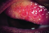
)
Fig. C14 'Cobblestones' papillae in severe vernal conjunctivitis
Millodot: Dictionary of Optometry and Visual Science, 7th edition. © 2009 Butterworth-Heinemann
con·junc·ti·vi·tis
(kŏn-jŭngk'ti-vī'tis)Inflammation of conjunctiva.
Medical Dictionary for the Dental Professions © Farlex 2012
Patient discussion about conjunctivitis
Q. What Causes Conjunctivitis? I woke up this morning with a red eye. My doctor said it's probably conjunctivitis. What causes this?
A. Red eye as a result of conjunctivitis is caused by hyperemia of the superficial blood vessels of the conjunctiva in the eye. This is usually caused due to either an allergic reaction, infection or some other trigger such as a foreign body that penetrated the eye. The blood vessels in the eye become engorged and therefore seen as red on the white sclera. This requires immediate medical examination in order to rule out emergency situations that can lead to permanent damage.
More discussions about conjunctivitisThis content is provided by iMedix and is subject to iMedix Terms. The Questions and Answers are not endorsed or recommended and are made available by patients, not doctors.