brain
[brān]that part of the central nervous system contained within the cranium, comprising the forebrain (prosencephalon), midbrain (mesencephalon), and hindbrain (rhombencephalon); it develops from the embryonic neural tube. The brain is a mass of soft, spongy, pinkish gray nerve tissue that weighs about 1.2 kg in a human being. It is connected at its base with the spinal cord, which is also part of the central nervous system. Called also encephalon. (See also color plates.)
The brain is made up of billions of nerve cells, intricately connected with each other. It contains nerve centers (groups of neurons and their connections) which control many involuntary functions, such as circulation, temperature regulation, and respiration, and interpret sensory impressions received from the eyes, ears, and other sense organs. Consciousness, emotion, thought, and reasoning are functions of the brain. It also contains centers or areas for associative memory which allow for recording, recalling, and making use of past experiences.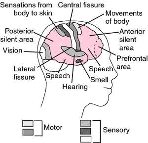
The brain is made up of billions of nerve cells, intricately connected with each other. It contains nerve centers (groups of neurons and their connections) which control many involuntary functions, such as circulation, temperature regulation, and respiration, and interpret sensory impressions received from the eyes, ears, and other sense organs. Consciousness, emotion, thought, and reasoning are functions of the brain. It also contains centers or areas for associative memory which allow for recording, recalling, and making use of past experiences.
Cerebrum. The largest and main portion of the brain, the cerebrum is made up of an outer coating, or cerebral cortex, consisting of gray matter, several cell layers deep, covering the cerebral hemispheres. The cortex is the thinking and reasoning brain, the intellect, as well as the part of the brain that receives information from the senses and directs the conscious movements of the body.
In appearance the cortex is rather like a relief map, with one very deep valley (longitudinal fissure) dividing it lengthwise into symmetrical halves, and each of the halves again divided by two major valleys and many shallower folds. The longitudinal fissure runs from the brow to the back of the head, and deep within it is a bed of matted white fibers, the corpus callosum, which connects the left and right cerebral hemispheres.
The major folds of the cortex divide each hemisphere into four sections or lobes: the occipital lobe at the back of the skull, the parietal lobe at the side, the frontal lobe at the forehead, and the temporal lobe at the temple.
The Senses. The major senses of vision and hearing have been well mapped in the cortex; the center for vision is at the back, in the occipital lobe, and the center for hearing is at the side, in the temporal lobe. Two other areas have been carefully explored, the sensory and motor areas for the body, which parallel each other along the fissure of Rolando. In the sensory strip are the brain cells that register all sensations, and in the motor strip are the nerves that control the voluntary muscles. In both, the parts of the body are represented in an orderly way.
It is in the sensory areas of the brain that all perception takes place. Here sweet and sour, hot and cold, and the form of an object held in the hand are recognized. Here are sorted out the sizes, colors, depth, and space relationships of what the eye sees, and the timbre, pitch, intensity, and harmony of what the ear hears. The significance of these perceptions is interpreted in the cortex and other parts of the brain. A face is not merely seen; it is recognized as familiar or interesting or attractive. Remembering takes place at the same time as perception, so that other faces seen in the past, or experiences linked to that face are called up. Emotions may also be stirred. For this type of association the cortex draws on other parts of the brain by way of the communicating network of nerves.
It is in the sensory areas of the brain that all perception takes place. Here sweet and sour, hot and cold, and the form of an object held in the hand are recognized. Here are sorted out the sizes, colors, depth, and space relationships of what the eye sees, and the timbre, pitch, intensity, and harmony of what the ear hears. The significance of these perceptions is interpreted in the cortex and other parts of the brain. A face is not merely seen; it is recognized as familiar or interesting or attractive. Remembering takes place at the same time as perception, so that other faces seen in the past, or experiences linked to that face are called up. Emotions may also be stirred. For this type of association the cortex draws on other parts of the brain by way of the communicating network of nerves.
Memory. In the temporal lobe, near the auditory area, is a center for memory. This center appears to be a storehouse where memories are filed. When this area alone is stimulated, a particular event, a piece of music, or an experience long forgotten or deeply buried is brought to the individual's mind, complete in every detail. This is a very mechanical type of memory; when the stimulation is removed the memory ends. When it is applied again, the memory begins again, not where it left off, but from the beginning.
Brainstem. This is the stemlike portion of the brain connecting the cerebral hemispheres with the spinal cord, and comprising midbrain, pons, and medulla oblongata. Some consider it to include the diencephalon.
Thalamus. This organ lies beneath the cortex, deep within the cerebral hemispheres. It is a relay station for body sensations, and integrates these sensations on their way to the cortex. The thalamus is an organ of crude consciousness and of sensations of rough contact and extreme temperatures, either hot or cold. It is principally here that pain is felt. In the thalamus, responses are of the all-or-nothing sort; even mild stimuli would be felt as acutely painful if they were not graded and modified by the cortex.
Hypothalamus. This organ lies below the thalamus, at the base of the cerebrum. It is small (no larger than a lump of sugar), but takes part in such vital activities as the ebb and flow of the body's fluids and the regulation of metabolism, blood sugar levels, and body temperature. It directs the body's many rhythms, including those of activity and rest, appetite and digestion, sexual desire, and menstrual and reproductive cycles. The hypothalamus is also the body's emotional brain. It is the integrating center of the autonomic nervous system, with its sympathetic and parasympathetic branches, and is located close to the pituitary gland.
Midbrain. Just below the thalamus is the short narrow pillar of the midbrain. This contains a center for visual reflexes, such as moving the head and eyes, as well as a sound-activated center, obsolete in humans, for pricking up the ears.
Medulla Oblongata. Below the midbrain is the medulla oblongata, the continuation upward of the spinal cord. In the medulla, the great trunk nerves, both motor and sensory, cross over, left to right and right to left, producing the puzzling phenomenon by which the left cerebral hemisphere controls the right half of the body, while the right hemisphere controls the left half of the body. This portion of the brain also contains the centers that activate the heart, blood vessels, and respiratory system.
Cerebellum. The cerebellum (Latin for “little brain”) is attached to the back of the brainstem, under the curve of the cerebrum. It is connected, by way of the midbrain, with the motor area of the cortex and with the spinal cord, as well as with the semicircular canals, the organs of balance. The function of the cerebellum is apparently to blend and coordinate motion of the various muscles involved in voluntary movements. It does not direct these movements; that is the function of the cortex. The cortex, however, operates in terms of movements, not of muscles. As a conscious function the cortex may, for example, direct the arm to pick up a glass of water; the cerebellum, which operates entirely below the level of consciousness, then translates this instruction into detailed actions by the 32 different muscles in the hand, plus several more in the arm and shoulder. When the cerebellum is injured, the patient's movements are jerky and uncoordinated.
Cranial Nerves. These are twelve nerves that arise within the skull. All but the olfactory nerve emerge from the brainstem. Most, with the important exception of the vagus nerve, serve the head and neck. See also cranial nerves.
Protection of the Brain. The brain is protected by the bony skull and by three layers of membranes, the meninges. Between the middle and inner layer is a space filled with cerebrospinal fluid, which serves as a shock absorber. The same system of membranes and fluid protects the spinal cord. The brain is protected from harmful substances in the bloodstream by a barrier called the blood-brain barrier, which keeps some of the substances out of the brain entirely and delays the entry of others for hours or even days after they have penetrated the rest of the body.

Projection areas of the brain.
brain abscess a localized suppurative lesion within the intracranial cavity; most cases are secondary to middle ear infections. Other causes include compound fracture of the skull with contamination of brain tissue, sinusitis, and infections of the face, lung or heart. Symptoms include fever, malaise, irritability, severe headache, convulsions, vomiting, and other signs of intracranial hypertension. Treatment consists of surgical removal of the infected area and administration of antibiotics.
brain death the irreversible cessation of all brain activity for an appropriate observation period, at least 24 hours, so that cardiopulmonary functions must be artificially maintained. A presidential commission in the USA accepted criteria for such a diagnosis, including cessation of all brain functions, including cerebral functions and brainstem (reflex) functions; irreversibility of the cessation; establishment of the cause of coma, sufficient to explain the loss of brain function; exclusion of possibility of recovery of brain function; and persistence of the cessation for an appropriate period of observation or trial of therapy. Complicating conditions must also be excluded. Called also irreversible coma.
brain scanning a nuclear medicine procedure for the detection of brain tumors, areas of stroke syndrome, abscesses, hematomas, and other intracranial lesions. A radiopharmaceutical, such as 99mTc-pertechnetate, is injected intravenously and is carried to the brain, where it localizes around any lesion that alters the blood-brain barrier. A scintillation camera makes an image of the distribution of radioactivity in which a lesion appears as a region of increased activity. Computed tomography brain scanning is an alternative procedure, which is more effective than radionuclide scans for the detection of some lesions.
brain tumor a neoplasm of the intracranial portion of the central nervous system. Any abnormal growth within the skull creates a special problem because it is in a confined space and will press on normal brain tissue and interfere with the functions of the body controlled by the affected parts. This is true whether the tumor itself is benign or malignant. Fortunately, the functions of certain areas of the brain are well known, and a disturbance of some specific function guides the clinician to the affected area. If diagnosed early, a benign tumor often can be removed surgically with a good chance of recovery. Malignant tumors are more difficult to remove. The causes of brain tumors are not known. They are not common, but they can occur at any age and in any part of the brain. Some originate in the brain itself, while others metastasize from a tumor in another part of the body.
The symptoms of brain tumor vary and depend on the location and size of the tumor. Headache together with nausea is sometimes the first sign. The headache can be generalized or localized in one part of the head, and the pain is usually intense. Vomiting can be significant if it is sudden and without nausea. Disturbances of vision, loss of coordination in movement, weakness, and stiffness on one side of the body are also possible symptoms. Loss of sight, hearing, taste, or smell may result from brain tumor. A tumor can also cause a distortion of any of these senses, such as seeing flashes at the sides of the field of vision, or smelling odors or hearing sounds that do not exist. It can affect the ability to speak clearly or to understand the speech of others. Varying degrees of weakness or paralysis in the arms or legs may appear. A tumor may cause convulsions. Changes in personality or mental ability are rare in cases of brain tumor. When such changes occur they may take the form of lapses of memory or absentmindedness, mental sluggishness, or loss of initiative.
The symptoms of brain tumor vary and depend on the location and size of the tumor. Headache together with nausea is sometimes the first sign. The headache can be generalized or localized in one part of the head, and the pain is usually intense. Vomiting can be significant if it is sudden and without nausea. Disturbances of vision, loss of coordination in movement, weakness, and stiffness on one side of the body are also possible symptoms. Loss of sight, hearing, taste, or smell may result from brain tumor. A tumor can also cause a distortion of any of these senses, such as seeing flashes at the sides of the field of vision, or smelling odors or hearing sounds that do not exist. It can affect the ability to speak clearly or to understand the speech of others. Varying degrees of weakness or paralysis in the arms or legs may appear. A tumor may cause convulsions. Changes in personality or mental ability are rare in cases of brain tumor. When such changes occur they may take the form of lapses of memory or absentmindedness, mental sluggishness, or loss of initiative.
wet brain brain edema.
Miller-Keane Encyclopedia and Dictionary of Medicine, Nursing, and Allied Health, Seventh Edition. © 2003 by Saunders, an imprint of Elsevier, Inc. All rights reserved.
Brain
(brān),Walter Russell, English physician, 1895-1966. See: Brain reflex.
brain
(brān), [TA]That part of the central nervous system contained within the cranium.
See also: encephalon. Compare: cerebrum, cerebellum.
See also: encephalon. Compare: cerebrum, cerebellum.
[A.S. braegen]
Farlex Partner Medical Dictionary © Farlex 2012
brain
(brān)n.
a. The portion of the vertebrate central nervous system that is enclosed within the cranium, continuous with the spinal cord, and composed of gray matter and white matter. It is the primary center for the regulation and control of bodily activities, receiving and interpreting sensory impulses, and transmitting information to the muscles and body organs. It is also the seat of consciousness, thought, memory, and emotion.
b. A functionally similar portion of the invertebrate nervous system.
The American Heritage® Medical Dictionary Copyright © 2007, 2004 by Houghton Mifflin Company. Published by Houghton Mifflin Company. All rights reserved.
brain
The epicentre of the central nervous system, which is located within the cranial vault and divided into the right and left hemispheres. The brain functions as a primary receiver, organiser and distributor of information for the body; it is the centre of thought and emotion, co-ordinates and controls bodily activities and interprets sensory visual, auditory, olfactory, tactile and other information.Segen's Medical Dictionary. © 2012 Farlex, Inc. All rights reserved.
watershed infarct
Neurology Infarction of a region peripheral to 2 arteries and susceptible to ischemia; WIs are often hemorrhagic, as restoration of the circulation allows blood to flow into damaged capillaries and 'leak' into the ischemic tissue Watershed infarctions-locations
Brain After internal carotid artery occlusion, causing vascular 'steal' phenomena, or between the anterior and middle cerebral arteries, which may be compromised in circle of Willis occlusions, often in a background of generalized atherosclerosis and as a possible complication of directed therapeutic embolization; cerebral perfusion may be impaired by cardiac arrest, pericardial tamponade and ex-sanguination
Large intestine At either the splenic flexure, the site of anastomosis between the inferior and superior mesenteric arteries, or at the rectum, a region supplied by peripheral irrigation from the inferior mesenteric artery and the hypogastric artery
McGraw-Hill Concise Dictionary of Modern Medicine. © 2002 by The McGraw-Hill Companies, Inc.
brain
(brān) [TA]That part of the central nervous system contained within the cranium.
See also: encephalon
Compare: cerebrum, cerebellum
See also: encephalon
Compare: cerebrum, cerebellum
[A.S. braegen]
Medical Dictionary for the Health Professions and Nursing © Farlex 2012
brain
The central organ of the body, to the maintenance, supply, transport and protection of which all the remainder of the body is dedicated. The brain contain more than 100 billion nerve cells with more than 1015 synapses. There are two main parts to the brain, the cerebrum and the cerebellum. The larger part, the cerebrum, initiates and coordinates all voluntary and most involuntary functions and is the seat of emotion, memory and intelligence. It is the medium by which all sensation, and the results of the mechanisms underlying all satisfaction, are conveyed to consciousness. It is essentially concerned with the collection, processing and storage of information, with the correlation of new data with stored data and with the organization and control of resulting responsive action. Response to stimulus is of the essence of brain function. Much is known, from the effects of disease and injury, of the localization of functions, in the brain, such as movement, sensation, vision, hearing, smell and speech. The location of areas responsible for registration and recall of memory is known, but the physical basis of memory storage remains obscure. Memory is not, like some other functions, located in a single definable area but is probably dispersed into all areas concerned with functions which may involve it. The cerebellum, the smaller part, is concerned mainly with the complex computations necessary to organize the muscle contractions needed to maintain the balance of the body and to allow walking and other movements. More than one tenth of the cardiac output is required to maintain brain function. See also BRAINSTEM.Collins Dictionary of Medicine © Robert M. Youngson 2004, 2005
)
Fig. 80 Brain . The vertebrate brain.
brain
the enlarged part of the CENTRAL NERVOUS SYSTEM beginning at the anterior end of bilaterally symmetrical animals (see BILATERAL SYMMETRY). The enlargement is associated with the aggregation of sense organs at the point which first contacts the changing environment. The brain, together with the rest of the central nervous system, coordinates the body functions. See also HEAD, CEPHALIZATION.Collins Dictionary of Biology, 3rd ed. © W. G. Hale, V. A. Saunders, J. P. Margham 2005
brain
(brān) [TA]That part of the central nervous system contained within the cranium.
[A.S. braegen]
Medical Dictionary for the Dental Professions © Farlex 2012
Patient discussion about brain
Q. What effect it will have in his brain………. hi all…………..whenever my bipolar son gets in to different episodes it makes me to think what effect it will have in his brain……….does it got anything to do with brain? But It didn’t strike me to discuss about this with my doctor….
A. after a while without treatment it's hard to stay without any kind of brain damage... the brain is a biological material which is affected by materials that ravage it over and over again. bipolar disorder can get worse, adding hallucinations and such.
Q. Does the brain recognize pain? How does the brain recognize pain.
A. First let us see the creation of the nerves which are assigned for different duties. There are roughly twenty different kinds of nerve endings in your skin that tell you if something is hot, cold, or painful. These nerve endings convert mechanical, thermal, or chemical energy into electrical signals that convey information to the brain and spinal cord - also known as the central nervous system or CNS. These signals travel to areas of your CNS where you perceive the stimuli as the painful sensations you actually feel - sensations such as searing, burning, pounding, or throbbing. Research suggests that the pain associated with fibromyalgia is caused by a "glitch" in the way the body processes pain. This glitch results in a hypersensitivity to stimuli that normally are not painful. According to the National Institute of Arthritis and Musculoskeletal and Skin Diseases (NIAMS), research has shown that people with fibromyalgia have reduced blood flow to parts of the brain.
Q. Is surfing the internet good for your brain? I am 72 and I just discovered computers and the internet at our library. I find myself fascinated by it and I spend hours in front of the computer. Is surfing the internet good for your brain?
A. This is a very current question that people ask and the answer is YES it is. A recent study showed that adults who surf the internet regularly engage larger parts of their brain when doing so compared to adults who rarely surf.
More discussions about brainThis content is provided by iMedix and is subject to iMedix Terms. The Questions and Answers are not endorsed or recommended and are made available by patients, not doctors.