aberration
[ab″er-a´shun]1. deviation from the normal or usual.
2. imperfect refraction or focalization of a lens.
chromatic aberration unequal refraction by a lens of light rays of different lengths passing through it, producing a blurred image and a display of colors.
dioptric aberration (spherical aberration) inability of a spherical lens to bring all rays of light to a single focus.
ventricular aberration aberrant ventricular conduction.
Miller-Keane Encyclopedia and Dictionary of Medicine, Nursing, and Allied Health, Seventh Edition. © 2003 by Saunders, an imprint of Elsevier, Inc. All rights reserved.
ab·er·ra·tion
(ab'er-ā'shŭn),1. Deviation from the usual or normal course or pattern.
See also: chromosome.
See also: chromosome.
2. Deviant development or growth.
See also: chromosome.
See also: chromosome.
[L. aberratio]
Farlex Partner Medical Dictionary © Farlex 2012
aberration
(ăb′ə-rā′shən)n.
1. A deviation from what is considered proper or normal.
2. Psychology A disorder or abnormal alteration in one's mental state.
3.
a. A defect of focus, such as blurring in an image.
b. An imperfect image caused by a physical defect in an optical element, as in a lens.
4. Genetics A deviation in the normal structure or number of chromosomes in an organism.
The American Heritage® Medical Dictionary Copyright © 2007, 2004 by Houghton Mifflin Company. Published by Houghton Mifflin Company. All rights reserved.
Aberration
A defect, deviation, or irregularityGenetics Chromosomal aberration
Ophthalmology Any error that results in image degradation. Such errors may be chromatic, spherical, or astigmatic chromatic, and may include distortion or curvature of field; these can result from design or execution, or both.
Physics
(1) Failure of an optical or electronic lens to produce an exact geometric—and chromatic—correlation between an object and its image
(2) In a video capture device or cathode-ray tube, a deviation in which the electrostatic or electromagnetic lens does not bring the electron beam to sharply focused points on the target or screen, or to correct geometric positions, as the beam is deflected.
Psychiatry Mental aberration
Zoology A term which, if used to denote a number of individuals within a species, unequivocally signifies infra-subspecies rank
Segen's Medical Dictionary. © 2012 Farlex, Inc. All rights reserved.
aberration
Medtalk A defect, deviation, or irregularity Psychiatry See Mental disorder.McGraw-Hill Concise Dictionary of Modern Medicine. © 2002 by The McGraw-Hill Companies, Inc.
ab·er·ra·tion
(ab'ĕr-ā'shŭn)1. Deviation from the usual or normal course or pattern.
2. Deviant development or growth.
See also: chromosome aberration
See also: chromosome aberration
[L. aberratio]
Medical Dictionary for the Health Professions and Nursing © Farlex 2012
aberration
A deviation from normal. The term derives from the Latin aberrare , to wander off. See also ABNORMAL.Collins Dictionary of Medicine © Robert M. Youngson 2004, 2005
aberration
An optical defect in which the rays from a point object do not form a perfect point after passing through an optical system. See oblique astigmatism; coma; curvature of field; distortion.
axial chromatic aberration See aberration, longitudinal chromatic.
lateral chromatic aberration Defect of an optical system (eye, lens, prism, etc.) in which the size of the image of a point object is extended by a coloured fringe, due to the unequal refraction of different wavelengths (dispersion). Syn. chromatic difference of magnification; transverse chromatic aberration (TCA). See dispersion; doublet.
longitudinal chromatic aberration (LCA) Defect of an optical system (eye, lens, prism, etc.) due to the unequal refraction of different wavelengths (dispersion) which results in an extended image along the optical axis. In the eye, blue rays are focused in front of the retina (by about 1 D) and red rays slightly behind the retina (0.25-0.5 D) when relaxed. When the eye is accommodated for a near target, blue rays tend to be focused near the retina and red rays are focused behind the retina (1 D), because of a lag of accommodation usually occurring when viewing near targets (Fig. A1). Syn. axial chromatic aberration. See chromoretinoscopy; chromostereopsis; constringence; dispersion; doublet; achromatizing lens; macular pigment; duochrome test.
monochromatic aberration Defect of an optical system (eye, lens, prism, etc.) occurring for a single wavelength of light. There are five such aberrations: spherical aberration, coma, curvature of field, oblique astigmatism and distortion. Syn. Seidel aberration.
negative aberration See spherical aberration.
oblique aberration Aberration induced by a point object off the optical axis of the system. These comprise coma, curvature of field, distortion and oblique astigmatism.
positive aberration See spherical aberration.
prism aberration Additional effects of a prism on light, in addition to the expected change in direction of light. These effects include different magnifications, curvature of field and chromatic aberration.
Seidel aberration See monochromatic aberration.
spherical aberration Defect of an optical system due to a variation in the focusing between peripheral and paraxial rays. The larger the pupil size, the greater the difference in focusing between the two rays. In the gaussian theory, the focus of the optical system is attributed to the paraxial rays. The distance, in dioptres, between the focus of the paraxial rays and the peripheral rays represents the amount of longitudinal spherical aberration of the system. When the peripheral rays are refracted more than the paraxial rays, the aberration is said to be positive or undercorrected. When the peripheral rays are refracted less than the paraxial rays the aberration is said to be negative or overcorrected. The relaxed human eye has a small amount of positive spherical aberration (up to 1 D for a pupil of 8 mm diameter) (Fig. A2). See caustic; aplanatic lens; gaussian theory.
transverse chromatic aberration See lateral chromatic aberration.
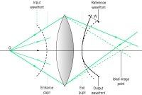
wavefront aberration The amount of deviation between an output wavefront emanating from an optical system and a conceptualized ideal (reference) wavefront. The specification of the deviation (or error) is usually fitted with a normalized Zernike expansion. The measurement of this aberration can be done subjectively or objectively (e.g. with an aberrometer based on the Hartmann-Shack principle). The method (called aberrometry) has been applied clinically to measure the aberrations displayed by optical systems, such as the eye, the eye with a correction, contact lenses (in vitro or in situ), intraocular lenses (in vitro or in situ), in corneal refractive surgery, cataract, etc. (Fig. A3). Syn. wave-front error.


axial chromatic aberration See aberration, longitudinal chromatic.
lateral chromatic aberration Defect of an optical system (eye, lens, prism, etc.) in which the size of the image of a point object is extended by a coloured fringe, due to the unequal refraction of different wavelengths (dispersion). Syn. chromatic difference of magnification; transverse chromatic aberration (TCA). See dispersion; doublet.
longitudinal chromatic aberration (LCA) Defect of an optical system (eye, lens, prism, etc.) due to the unequal refraction of different wavelengths (dispersion) which results in an extended image along the optical axis. In the eye, blue rays are focused in front of the retina (by about 1 D) and red rays slightly behind the retina (0.25-0.5 D) when relaxed. When the eye is accommodated for a near target, blue rays tend to be focused near the retina and red rays are focused behind the retina (1 D), because of a lag of accommodation usually occurring when viewing near targets (Fig. A1). Syn. axial chromatic aberration. See chromoretinoscopy; chromostereopsis; constringence; dispersion; doublet; achromatizing lens; macular pigment; duochrome test.
monochromatic aberration Defect of an optical system (eye, lens, prism, etc.) occurring for a single wavelength of light. There are five such aberrations: spherical aberration, coma, curvature of field, oblique astigmatism and distortion. Syn. Seidel aberration.
negative aberration See spherical aberration.
oblique aberration Aberration induced by a point object off the optical axis of the system. These comprise coma, curvature of field, distortion and oblique astigmatism.
positive aberration See spherical aberration.
prism aberration Additional effects of a prism on light, in addition to the expected change in direction of light. These effects include different magnifications, curvature of field and chromatic aberration.
Seidel aberration See monochromatic aberration.
spherical aberration Defect of an optical system due to a variation in the focusing between peripheral and paraxial rays. The larger the pupil size, the greater the difference in focusing between the two rays. In the gaussian theory, the focus of the optical system is attributed to the paraxial rays. The distance, in dioptres, between the focus of the paraxial rays and the peripheral rays represents the amount of longitudinal spherical aberration of the system. When the peripheral rays are refracted more than the paraxial rays, the aberration is said to be positive or undercorrected. When the peripheral rays are refracted less than the paraxial rays the aberration is said to be negative or overcorrected. The relaxed human eye has a small amount of positive spherical aberration (up to 1 D for a pupil of 8 mm diameter) (Fig. A2). See caustic; aplanatic lens; gaussian theory.
transverse chromatic aberration See lateral chromatic aberration.
wavefront aberration The amount of deviation between an output wavefront emanating from an optical system and a conceptualized ideal (reference) wavefront. The specification of the deviation (or error) is usually fitted with a normalized Zernike expansion. The measurement of this aberration can be done subjectively or objectively (e.g. with an aberrometer based on the Hartmann-Shack principle). The method (called aberrometry) has been applied clinically to measure the aberrations displayed by optical systems, such as the eye, the eye with a correction, contact lenses (in vitro or in situ), intraocular lenses (in vitro or in situ), in corneal refractive surgery, cataract, etc. (Fig. A3). Syn. wave-front error.
)
Fig. A1 Longitudinal chromatic aberration of the eye
)
Fig. A2 Spherical aberration of the eye. Two parallel rays coming from infinity are focused, one at F′, the secondary focal point corresponding to paraxial rays and the other peripheral ray in front or behind F′, depending on the type of spherical aberration
)
Fig. A3 An input spherical wavefront of light is centred on object O. After emerging from a lens affected by monochromatic aberration, it is no longer spherical and the image-forming rays do not meet in the single ideal image point (the paraxial image)
| Table A1 Aberrations of the eye | |||||||
| A | Chromatic aberrations: | ||||||
| longitudinal (or axial): chromatic difference of focus | |||||||
| transverse (or lateral): chromatic difference of magnification | |||||||
| B | Monochromatic aberrations (or Seidel aberrations): | ||||||
| Type | direction | stimulus | |||||
| 1. spherical aberration | longitudinal transverse | light beam passing through large pupil | |||||
| 2. coma | transverse | points objects off the optical axis | |||||
| 3. oblique astigmatism | longitudinal | points objects off the optical axis | |||||
| 4. curvature | longitudinal | extended objects of field | |||||
| 5. distortion | transverse | extended objects | |||||
| 6. wavefront aberration | transverse | extended objects | |||||
Millodot: Dictionary of Optometry and Visual Science, 7th edition. © 2009 Butterworth-Heinemann
ab·er·ra·tion
(ab'ĕr-ā'shŭn)1. Deviation from the usual or normal course or pattern.
2. Deviant development or growth.
See also: chromosome
See also: chromosome
[L. aberratio]
Medical Dictionary for the Dental Professions © Farlex 2012